
Page 25 - Journal of Structural Heart Disease Volume 3, Issue 2
P. 25
51
Case Report
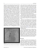
angioplasties were performed in the narrowed SVC using a 5 × 20 mm Sterling balloon (Boston Scien- ti c, Marlborough, Massachusetts) over a .018 Plati- num Plus wire (Boston Scienti c, Marlborough, Mas- sachusetts), but a tight waist persisted at 14 ATM of pressure (Figure 2). Dilations were then performed with a higher pressure 5 × 20 mm Dorado balloon (Bard Medical, Covington, Georgia) over the same .018 wire, which resulted in resolution of the tight waist at 24 ATM. However, on repeat angiography, although the SVC showed improved ow, there was signi cant vascular recoil and residual obstruction necessitating stent implantation. In anticipation of stent implantation, the central venous line from the right subclavian vein that crossed the SVC was pulled out of the SVC and into the left innominate vein using a snare catheter introduced from the he- miazygos vein. An 8F long sheath was placed in the SVC from the femoral vein over a .035 Amplatz Super Sti wire (Boston Scienti c, Marlborough, Massachu- setts), and a Palmaz Genesis 1910 XD stent (Cordis, Fremont, California) mounted on a 6 × 20 mm Do- rado balloon was deployed in the SVC and further dilated with a 7 × 20 mm Dorado balloon at 22 ATM. Afterward, there was a 1 mmHg gradient across the
Figure 2. Balloon dilation shows a signi cant residual waist using a 5-mm Sterling balloon at 14 ATM of pressure. The stenosis ulti- mately required 22-24 ATM of pressure to resolve at full in ation.
stent, and the SVC was widely patent, measuring 6 mm. At the conclusion of the procedure, there was excellent ow through the well-positioned stent, and the veins no longer decompressed through the hemiazygos system; rather, all venous ow en- tered the heart briskly through the SVC (Figure 3). Of note, the patient required balloon venoplasty of the right iliofemoral vein due to narrowing, likely from a previous line placement. After venoplasty, a PICC line was placed in the right femoral vein so that the tunneled subclavian line could be removed.
The patient was transitioned off octreotide over the following 4 days. Medium-chain triglyceride for- mula was replaced with breast milk, and no recur- rence of chylous pleural effusion was observed. The chest tube was removed on post-catheterization day 6.
To preserve and rehabilitate the obstructed right femoral vein, repeat catheterization was performed 11 days after initial catheterization, at which time the right femoral vein PICC was removed and an- gioplasty was performed to maintain the patency of the right iliofemoral vein. Repeat angiography of the SVC stent showed continued unobstructed ow across the SVC stent with no short-term re- current narrowing (Figure 4). The patient was dis- charged 2 days later on low-dose aspirin for stent endothelialization.
At the most recent follow-up 5 months after dis- charge, echocardiogram showed excellent ow through a stably positioned SVC stent with a mean Doppler gradient of 3.8 mmHg. The dilated hemia- zygous vein was no longer visualized. No recurrent pleural e usions were noted on follow-up chest ra- diography. The patient will require periodic repeat catheterizations with angioplasty of the SVC stent for somatic growth until the vessel reaches adult size.
Discussion
Chylothorax is a known cause of respiratory com- promise and appears at an increased frequency in patients with congenital heart disease, in particular following intrathoracic surgical intervention. Chylot- horax is most commonly due to incidental damage to the thoracic duct during a surgical procedure or, alternatively, to venous stenosis/obstruction causing
Harrison, D. J. et al.
Intravascular Stent Implantation for Chylothorax
