
Page 26 - Journal of Structural Heart Disease Volume 3, Issue 2
P. 26
Case Report 52
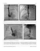
Figure 3. Panel A. After implantation of a PG1910XD stent in the superior vena cava and high-pressure dilation to 7 mm, there was in- creased forward ow across the stent into the right atrium and no retrograde ow in the innominate vein or decompression through collateral veins. Note that the right subclavian central venous line was repositioned in the left innominate vein using a snare catheter from the hemiazygos vein (snaring not pictured). Panel B. Lateral view after stent dilation.
Figure 4. Panel A. Two weeks after the initial procedure, there was continued unobstructed ow across the stent and no short-term recurrent stenosis. Panel B. Lateral view of 2-week follow-up.
increased pressure in the thoracic duct [5]. The most common site for this type of obstruction is the innomi- nate vein [6], but as demonstrated in this case, the SVC
must also be considered as a potential site of vascular compromise. The most common causes of SVC steno- sis or obstruction leading to chylothorax in children
Journal of Structural Heart Disease, April 2017
Volume 3, Issue 2:49-54
