
Page 29 - Journal of Structural Heart Disease Volume 3, Issue 4
P. 29
Case Report 116
ABCD
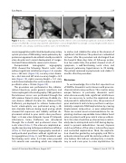
Figure 1. Cardiac computerized tomographic angiography showed a brotic aortic valve without signi cant calci cations (Panel A), annular area (Panel B), coronary sinus diameter and left main coronary height (Panel C), and right coronary height and the distance between the aortic annulus and mitral prosthetic valve = 10 mm (Panel D).
severe regurgitation with calculated pulmonary artery systolic pressure of 100 mmHg. Severe pulmonary hy- pertension appeared to be related to both prior mitral valve disorder and a recent development of conges- tive heart failure related to severe aortic stenosis.
Computerized tomographic angiography (CTA) showed the following: brotic aortic valve without signi cant calci cation (Figure 1A), annular area = 365 mm2 (Figure 1B), coronary sinus diame- ter = 26.5 mm and left main coronary height =12.9 mm (Figure 1C), right coronary height = 12.1 mm, and distance between the aortic annulus and mitral prosthetic valve = 10 mm (Figure 1D).
The procedure was performed in the catheter- ization laboratory under general anesthesia and transesophageal echocardiography ( TEE) guidance. Percutaneous access was performed through the left common femoral artery with two ProGlide clo- sure devices (Abbott Vascular Inc., Redwood City, California) pre-deployed to achieve homeostasis. Aortic valvuloplasty was performed using a 20- mm diameter balloon during rapid pacing, which showed full expansion and stability without dis- placement related to prior mitral valve prosthesis. Next, a 23-mm valve (Edwards Sapien XT, Edwards Lifesciences, Irvine, California) was introduced through the e-sheath and positioned across the aortic valve (Video 1). The valve was implanted us- ing the rapid pacing technique and balloon in ation ( Video 2). Post-procedural angiography revealed a well-positioned prosthesis without signi cant aor- tic regurgitation (Video 3), and TEE revealed a well- functioning aortic valve. A 23-mm valve provided 14% annular area oversizing, which was adequate
to anchor and stabilize the valve in the absence of signi cant calci cation. The patient was ambulated six hours after the procedure and discharged from the hospital three days later. At follow-up evalua- tion two weeks later, the patient showed a lack of symptoms, a well-functioning aortic valve, and improved pulmonary hypertension to 70 mmHg. The patient remained asymptomatic and stable 15 months after the procedure.
Discussion
To our knowledge, this is the first reported case of TAVR for rheumatic aortic stenosis with prior me- chanical mitral valve prosthesis. This case has many unique features. In particular, rheumatic aortic valve disease usually lacks significant calcifications that are necessary to anchor the implanted valve and prevent its embolization. Also, the presence of prior mechanical mitral valve prosthesis could po- tentially complicate TAVR implantation by causing displacement, malposition, or even embolization. There are several usual recommendations for im- planting a balloon-expandable transcatheter heart valve in patients with prior mitral valve prosthesis. First, the valve should be positioned more ventric- ularly due to anticipated aortic shift during deploy- ment to prevent embolization. Second, stepwise and slow inflation of the valve is advised for proper and controlled implantation. Third, the implanta- tion should be guided by aortography and TEE to evaluate interaction with the mitral prosthesis. We also believe that balloon valvuloplasty is manda- tory in such cases prior to valve implantation for
Journal of Structural Heart Disease, August 2017
Volume 3, Issue 4:115-118
