
Page 30 - Journal of Structural Heart Disease Volume 3, Issue 4
P. 30
117
Case Report
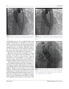
Video 1. Valve positioning prior to implantation. View supple- mental video at https://doi.org/10.12945/j.jshd.2017.015.16. vid.01.
the following reasons. First, an inflated balloon can confirm full expansion of the fibrotic aortic valve leaflets. Second, balloon stability during inflation can indicate good anchoring, suggesting that the implanted valve is less likely to embolize. Third, it can allow examination of the interaction between the aortic valve and mitral prosthesis that could potentially cause displacement or malposition.
Rheumatic valve disease usually inflicts younger patients, and surgical valve replacement is the procedure of choice for these patients. However, the presence of comorbidities (i.e., liver cirrhosis) and prior mitral valve prosthesis makes TAVR a po- tentially excellent solution for severe aortic stenosis.
Review of the literature revealed several case re- ports of TAVR in rheumatic heart disease. Bilge et al. described two patients with rheumatic aortic steno- sis and high surgical risk who ultimately underwent TAVR [9]. Akujuo et al. presented a case in which TAVR was combined with transcatheter mitral valve implantation in a patient with both aortic and mi- tral valves a ected by rheumatic valve stenosis [10]. Chainani et al. reported a case of left main percuta- neous coronary intervention and TAVR in a patient with rheumatic heart disease and porcelain aorta
Video 2. Aortic valve implantation during rapid pacing. View sup- plemental video at https://doi.org/10.12945/j.jshd.2017.015.16. vid.02.
Video 3. Post trans-catheter aortic valve replacement angi- ography showing well-positioned valve without signi cant aortic regurgitation. View supplemental video at https://doi. org/10.12945/j.jshd.2017.015.16.vid.03.
Alhaddad, I.A.
TAVR for Rheumatic Aortic Stenosis
