
Page 38 - Journal of Structural Heart Disease Volume 3, Issue 4
P. 38
125 Case Report
Video 5. Selective left coronary angiography following Melody TPV implantation at steep caudal projections showing no signif- icant coronary arterial narrowing after PPVI. The pressure wire was positioned in the distal LAD. View supplemental video at https://doi.org/10.12945/j.jshd.2017.017.17.vid.05.
Video 6. Main pulmonary artery angiography following Melo- dy TPV implantation demonstrating trivial central valvar regur- gitation with a catheter through the valve and no paravalvar leak. View supplemental video at https://doi.org/10.12945/j. jshd.2017.017.17.vid.06.
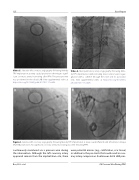
Figure 6. Selective left coronary angiography following Melody TPV implantation at steep caudal (Panel A) and left anterior oblique (Panel B) projections. No signi cant coronary arterial narrowing was seen following PPVI.
continuously monitored via a pressure wire during the intervention. Although the left coronary artery appeared remote from the implantation site, there
were potential masses (e.g., calci cation, scar tissue) in addition to the pre-stents that could result in coro- nary artery compression. Continuous distal LAD pres-
Boe, B. A. et al.
CA Pressure Wire During PPVI
