
Page 37 - Journal of Structural Heart Disease Volume 3, Issue 4
P. 37
Case Report 124
B
C
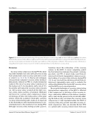
Figure 5. Continuous pressure monitoring of the proximal left main coronary artery (red) and distal LAD artery (yellow). At baseline (Panel A), the pressure in the LAD was slightly lower than the left main pressure. With balloon sizing of the RVOT (Panel B), both pres- sures decreased equally and returned to baseline upon de ation of the sizing balloon (Panel C). The lateral projection of the balloon sizing (Panel D) showed a balloon waist of 19.8 mm in diameter inferior to the left main coronary artery.
Discussion
Coronary artery compression during PPVI has been reported in multiple case series with outcomes varying from symptomatic acute coronary syndrome to cardi- ac arrest [5, 6, 7, 8, 9, 10, 11]. Morray and colleagues retrospectively evaluated coronary artery testing in 404 patients referred for PPVI in a multi-institutional study [4]. The risk of coronary artery compression in the study was 4.7% for all patients, with a risk of 71% for patients with abnormal coronary artery anatomy (i.e., left coronary artery arising from the right coro- nary artery, status post-Ross procedure). Additional risk factors for coronary artery compression includ- ed tetralogy of Fallot and transposition of the great arteries. Given this potential catastrophic outcome, coronary arterial testing with a balloon of equal size to the intended pre-stent implantation balloon is rec- ommended and is the standard of care during PPVI. Although coronary artery testing provides good in-
formation about the relationship of the coronary arteries to the RVOT, it cannot completely predict the nal interaction between the patient’s anatomy, pre-stents, and TPV. A recent study used three-di- mensional rotational angiography to improve our un- derstanding of coronary artery anatomy during PPVI [12]. In our case, both two-dimensional and three-di- mensional angiography were used to evaluate the relationship between the stented left coronary artery and the RVOT prior to any intervention.
The accepted technique of coronary artery testing during balloon angioplasty of the RVOT is e ective if the compression or distortion of the coronary ar- tery is relieved by de ation of the balloon. However, with a stent already present in the at-risk LAD, de at- ing the balloon may not relieve the coronary artery compression. A stent implanted within the RVOT or coronary artery may prevent reversible coronary ar- terial compression. Here, we describe the rst PPVI in a patient with coronary arterial stents that were
Journal of Structural Heart Disease, August 2017
Volume 3, Issue 4:119-127
