
Page 36 - Journal of Structural Heart Disease Volume 3, Issue 4
P. 36
123
Case Report
Video 4. Selective left coronary angiography showing the tip of the pressure wire positioned in the LAD distal to the previ- ously placed stents. View supplemental video at https://doi. org/10.12945/j.jshd.2017.017.17.vid.04.
Milpitas, California) delivered on 22 mm × 3.5 cm BIB balloons (NuMED, Inc., Hopkinton, New York) without a signi cant pressure di erence across the LAD stent. The stents overlapped at the RVOT narrowing, which was the intended landing zone for the valve. A PB1018 Melody Transcatheter Pul- monary Valve® (TPV) (Medtronic, Minneapolis, Minnesota) was delivered via a 22-mm Ensemble® Transcatheter Valve Delivery System (Medtronic) coaxially within the Palmaz stents. Distal LAD cor- onary arterial pressure remained stable during the RVOT intervention. Repeat selective left coronary arterial angiography was performed following Mel- ody TPV implantation, which showed no compres- sion of the coronary artery (Video 5, Figure 6). The pressure wire and JL guide catheter were then re- moved. Intracardiac echocardiographic evaluation of the Melody TPV revealed trivial central valvar regurgitation and no paravalvar leak (Video 6). The patient tolerated the procedure without complica- tion and was discharged home on Aspirin 325 mg and Clopidogrel 75 mg daily.
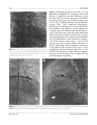
Figure 4. Anteroposterior (Panel A) and lateral (Panel B) projections of selective left coronary angiography showing the pressure wire (arrow) positioned in the distal LAD artery distal to the previously placed drug-eluting coronary stents.
Boe, B. A. et al. CA Pressure Wire During PPVI
