
Page 34 - Journal of Structural Heart Disease Volume 3, Issue 4
P. 34
121
Case Report
Video 2. Three-dimensional rotational reconstruction of the right ventricular out ow tract (yellow) and left coronary artery (blue) from dual injection three-dimensional rotational angi- ography. The left main coronary artery ran posterior to the out- ow tract and was superior to the intended valve implantation site at the level of the native pulmonary valve annulus. The LAD coursed leftward and posterior to the right ventricular out ow tract. View supplemental video at https://doi.org/10.12945/j. jshd.2017.017.17.vid.02.
balloon sizing of the RVOT with a low-pressure bal- loon. Given the risk of coronary stent compression with balloon angioplasty, stent implantation, and PPVI within the RVOT, we decided to continuously monitor distal LAD pressure during the interven- tion. This plan was discussed with our adult inter- ventional colleagues, who were prepared to provide support as needed.
A 6-F JL-4 Guiding catheter (Cordis®, Milpitas, California) was used to cannulate the left main cor- onary artery, and a 0.014” PrimeWire Prestige® PLUS pressure guide wire (Volcano Co., San Diego, Cal- ifornia) was positioned in the distal LAD (Video 4, Figure 4). The measured pressure from the pressure wire was calibrated with the proximal coronary ar- terial pressure measured from the guide catheter. A 2.5 × 15 mm Maverick coronary balloon (Boston Scientific Co., Marlborough, Massachusetts) was prepped and ready for use in the case of a crush injury to a coronary artery stent during RVOT intervention.
Sizing of the RVOT was performed using a 25 mm × 4 cm Tyshak II balloon (B. Braun Medical Inc., Bethlehem, Pennsylvania) in ated to 1 atm. Selec- tive left coronary angiography and continuous LAD pressure monitoring was performed during balloon sizing of the RVOT. Both the proximal left coronary
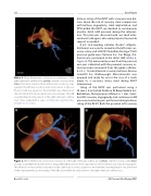
Figure 2. Three-dimensional rotational reconstruction of the right ventricular out ow tract (yellow) and left coronary artery (blue) in steep caudal (Panel A) and left anterior oblique (Panel B) projections from a dual injection three-dimensional rotational angiogram. The left main coronary artery ran posterior to the out ow tract and was superior to the intended valve implantation site at the level of the native pulmonary valve annulus. The LAD coursed leftward and posterior to the right ventricular out ow tract.
Boe, B. A. et al. CA Pressure Wire During PPVI
