
Page 33 - Journal of Structural Heart Disease Volume 3, Issue 4
P. 33
Case Report 120
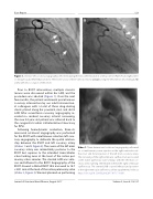
Figure 1. Selective left coronary angiography performed during the rst catheterization in anteroposterior (Panel A) and right anteri- or oblique/caudal (Panel B) projections. There were a total of three stenotic lesions (arrows) along the left anterior descending (LAD) artery with near occlusion of the vessel.
Prior to RVOT intervention, multiple stenotic lesions were discovered within her LAD, and the procedure was aborted (Figure 1). Over the next few months, the patient underwent percutaneous coronary intervention by our adult intervention- al colleagues with a total of three drug-eluting stents placed along her proximal, mid, and distal LAD. After surveillance coronary angiography re- vealed no residual coronary arterial narrowing, the now 54-year-old patient was referred back to the congenital cardiac catheterization laboratory for PPVI.
Following hemodynamic evaluation, three-di- mensional rotational angiography was performed for the RVOT with simultaneous selective left coro- nary angiography to delineate the spatial relation- ship between the RVOT and left coronary artery (Videos 1 and 2 Figure 2). The course of the left main coronary artery was immediately posterior to the RVOT but superior to the intended transcatheter valve landing zone at the level of the native pul- monary valve annulus. The stented LAD was ante- rior and leftward to the RVOT. Angiography of the RVOT showed a dilated RVOT that narrowed to 16.7 × 20.1 mm at the level of the native pulmonary valve (Video 3, Figure 3). We next planned on performing
Video 1. Three-dimensional rotational angiography performed via a simultaneous power injection in the right ventricular out- ow tract and hand injection of the left main coronary artery. The narrowing of the right ventricular out ow tract was located at the native pulmonary valve annulus with the left main cor- onary artery running immediately behind the right ventricular out ow tract. The stented LAD coursed leftward and posterior to the right ventricular out ow tract. View supplemental video at https://doi.org/10.12945/j.jshd.2017.017.17.vid.01.
Journal of Structural Heart Disease, August 2017
Volume 3, Issue 4:119-127
