
Page 35 - Journal of Structural Heart Disease Volume 3, Issue 4
P. 35
Case Report 122
Video 3. Angiography of the right ventricular out ow tract in the anteroposterior (Panel A) and lateral (Panel B) projections. The narrowing of the right ventricular out ow tract occurred at the native pulmonary valve annulus. View supplemental video at https:// doi.org/10.12945/j.jshd.2017.017.17.vid.03A and https://doi.org/10.12945/j.jshd.2017.017.17.vid.03B.
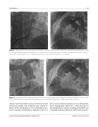
Figure 3. Angiography of the right ventricular out ow tract in the anteroposterior (Panel A) and lateral (Panel B) projections with measurements. The narrowing at the native pulmonary valve annulus measured 17.1 × 20.1 mm in both projections.
arterial and distal LAD coronary arterial pressures decreased equally and simultaneously during in- ation of the sizing balloon and completely recov- ered to baseline with balloon de ation (Figure 5).
No coronary arterial compression was demonstrat- ed by angiography. There was a 19.8-mm waist on the sizing balloon, and pre-stenting of the RVOT was completed with two Palmaz 3110 XL stents (Cordis®,
Journal of Structural Heart Disease, August 2017
Volume 3, Issue 4:119-127
