
Page 27 - Journal of Structural Heart Disease Volume 4, Issue 1
P. 27
Case Report
18
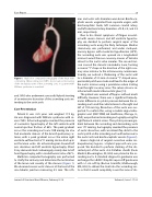
Figure 1.
mus and aortic arch diameters were 6 mm. Brachioce- phalic vessels originated from separate origins, with brachocephalic trunk, left common carotid artery, and left subclavial artery diameters of 3.0, 2.8, and 3.8 mm, respectively.
Due to his clinical symptoms of fatigue associat- ed with severe stenosis and left ventricle hypertro- phy, we decided to perform surgical repair of the ascending aorta using the Doty technique. Median sternotomy was performed, and under cardiopul- monary bypass with moderate hypothermia (28°C), the ascending aorta was opened via a longitudinal incision toward the non-coronary sinuses of Valsalva almost to the aortic valve annulus. The second inci- sion crossed the stenotic sinotubular zone, forming a reverse “Y” shape in the direction of the right coro- nary sinus anterior to the intracoronary commissure. Visually, we noticed a thickening of the aortic wall to a diameter of 3.5 mm. A reverse “Y”-shaped xeno- pericardial patch was made and xed to the edges of the aortic incision using a premilene suture starting from the right coronary sinus. The suture site was re- inforced with medical hemostatic glue [14].
The patient was weaned o bypass without much di culty. However, there was a signi cant hemody- namic di erence in systolic pressure between the as- cending aorta and the radial arteries to the right and left of 110 mm Hg. Dissection of the aorta was sus- pected. To con rm this, using a mobile angiocardio- graphic unit (OEC 9900, GE Healthcare, Chicago, IL, USA), we performed ascending aortography using the right femoral arterial access. The systolic pressure gra- dient between the ascending and descending aorta was 177 mm Hg. Aortography revealed the presence of aortic dissection, with an intimal ap distal to the aortic patch on the ascending aorta with extension to the aortic arch and brachiocephalic vessels (Figure 2).
Given a high risk of surgical correction under car- diopulmonary bypass, a hybrid approach was pro- posed. We decided to perform stenting of the dis- sected part of the aorta. A 6-F Mullins sheath (Cook Medical, Bloomington, IN, USA) was placed in the as- cending aorta. A standard diagnostic guidewire was exchanged for a 0.035” Amplatz super-sti guide wire with a 1-cm soft tip (Boston Scienti c, Marlborough, MA, USA) to deliver the stent into the ascending aor- ta so that it would completely cover the zone of dis-
Multi-slice computed tomography of the heart with contrasting. 3D-reconstruction (VRT). A pronounced narrowing of the sinotubular zone of the ascending aorta in a patient with Williams syndrome is visualized.
and SVAS who underwent successful hybrid stenting of an extensive dissection of the ascending aorta ex- tending to the aortic arch.
Case Presentation
Patient K. was 2.25 years and weighed 11.9 kg. He was diagnosed with Williams syndrome with se- vere SVAS. Echocardiography revealed the presence of concentric hypertrophy of the left ventricle with normal ejection fraction of 68%. The peak gradient across the ascending aorta was 100 mmHg. He also had moderate stenosis of the branch pulmonary ar- teries, with a peak gradient across the entire right ventricle out ow tract of 11 mmHg. He also had a pat- ent foramen ovale. His echocardiogram showed left axis deviation and left ventricle hypertrophy. Chest X-ray demonstrated cardiomegaly, mainly due to left heart hypertrophy, with a cardiothoracic ratio of 67%.
Multislice computed tomography was performed to clarify the anatomy and determine the localization of the lesion and severity of the stenosis (Figure 1). We observed severe aortic stenosis at the level of the sino-tubular junction measuring 4.5 mm. The isth-
Journal of Structural Heart Disease, February 2018
Volume 4, Issue 1:17-20
