
Page 28 - Journal of Structural Heart Disease Volume 4, Issue 1
P. 28
19
Case Report
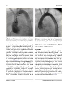
Figure 2. Aortography in the left oblique projection. There is a dissection of the ascending section and the aortic arch (indicat- ed with arrows). The dissection extends to the mouth of the bra- chiocephalic trunk and the left common carotid artery.
section to the point of origin of the brachiocephalic vessels. A 18-mm Valeo stent (Bard, Murray Hill, NJ, USA) was attached to a 8-mm balloon deployed to 10 atm. After stent implantation, however, there was no signi cant change in systolic pressure gradient. Therefore, a second 26-mm Valeo stent on a 8-mm balloon was implanted covering the whole surface of the aortic arch. Repeat aortography revealed proper implantation of both stents covering the entire zone of dissection. Patency of the brachiocephalic arteries was preserved (Figure 3). Invasive pressure measure- ment after stent implantation showed minimal resid- ual systolic pressure of 15 mm Hg. The child left the operating room on adrenaline at a dose of 0.1 μg/kg/ min.
The child was extubated after 20 hours. Anticoag- ulant therapy was initiated using heparin at a rate of 200 U/kg/day for 3 days followed by aspirin at a dose of 50 mg/day. The patient was discharged home on postoperative day 13. Unfortunately, the parents re- fused postoperative follow-up consultation in our
Figure 3. Aortography in the left oblique projection. After the implantation of the two Valeo stents into the ascending aorta and the aortic arch, there are no signs of aortic narrowing and dissection, the brachiocephalic vessels are completely passable.
center due to a stated good clinical status of their child and distant place of residence.
Discussion
Surgical correction of SVAS in patients with Wil- liams syndrome is safe and e ective. However, a rare acute complication of dissection of the ascending aorta can lead to an unfavorable prognosis. There are no clear recommendations or algorithms for action in cases of dissection. In adult patients with acute dis- section, prosthetic material can be used to repair the dissection. In children, however, this is more di cult and sometimes not feasible. Therefore, in such cases, using endovascular techniques to eliminate acute aortic dissection is potentially a promising solution. We did not nd descriptions of similar clinical cases in young children in the literature. Available reports discussed planned endovascular and hybrid inter- ventions to eliminate residual stenosis of the ascend- ing aorta and aortic arch after previously performed reconstructive surgical interventions [15-16].
Pursanov M. G. et al.
Stenting of the Aortic Dissection in Children
