
Page 22 - Journal of Structural Heart Disease Volume 4, Issue 2
P. 22
43 Case Report
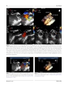
Figure 1. Transesophageal echocardiography (TEE) of transcatheter aortic valve replacement (TAVR) in D-transposition of the great arteries (D-TGA) status post arterial switch with a valve sparing root repair using a hemashield graft and repair of severe aortic regur- gitation using a 26-mm Edwards Sapien-3 valve. Panel A. TEE two-dimensional (2D) long axis view of the aortic valve demonstrating left coronary cusp prolapse and severe regurgitation. Panel B. TEE three-dimensional (3D) long axis view of the aortic valve demon- strating left coronary cusp prolapse and aberrant chord in the left ventricular out ow tract. Panel C. TEE 2D long axis view of the aortic valve status post deployment of a 26-mm Sapien-3 valve with no residual regurgitation and trace perivalvular regurgitation. Panel D. TEE 2D short axis view of the aortic valve status post deployment of a 26-mm Sapien-3 valve with no residual regurgitation and trace perivalvular regurgitation.
Video 1. TEE 2D long axis view of the aortic valve with color com- pare showing the prolapsing aortic valve lea et and severe regur- gitation. View supplemental video at https://doi.org/10.12945/j. jshd.2018.042.17.vid.01.
Video 2. TEE 3D view of the aortic valve prolapse and aberrant chord. View supplemental video at https://doi.org/10.12945/j. jshd.2018.042.17.vid.02.
Ghobrial J. et al.
TAVR in TGA
