
Page 24 - Journal of Structural Heart Disease Volume 4, Issue 2
P. 24
45
Case Report
Procedure
Pre-procedural electrocardiography-gated cardiac computed tomographic angiography (CTA) showed the valve annular dimensions to be 21 × 20 × 23 mm, adequate clearance of the coronary ostia to the annu- lus, and no aortic valve or ascending aortic calci ca- tion. Abdominal/pelvic CTA demonstrated adequate femoral arterial size for a transfemoral valve replace- ment approach without signi cant tortuosity.
The procedure was performed under general an- esthesia. Intra-procedural transesophageal echocar- diography (TEE) showed severe eccentric aortic re- gurgitation due to malcoaptation of the aortic valve lea ets and prominent prolapse of the left coronary cusp lea et (Figure 1; Videos 1, 2, 3, and 4). There was a false chord extending from the anterior mitral valve lea et to the left ventricular out ow tract without functional obstruction (Video 2). The aortic valve an- nulus measured 20 × 20 × 21 mm by TEE, mostly con- sistent with the CTA annular dimensions. Aortic root angiograms (Figure 2; Video 5) showed severe AR and patent re-implanted coronary arteries without evi- dence of atherosclerotic disease with a distance from the valve level of 27–30 mm and minimal concern for coronary obstruction. A 23 mm × 4 cm balloon was used to perform balloon sizing with rapid ventricu- lar pacing at 180 bpm; the waist on the balloon was noted to be 21 mm. A 26-mm Edwards Sapien-3 valve (Edwards LifeSciences, Irvine, CA, USA) was prepped and mounted onto the Commander delivery sys- tem in the usual manner and advanced into the 14-F sheath, and the valve was assembled in the descend- ing aorta. Once across the valve, aortic angiography was performed to ensure appropriate position of the Sapien valve. Rapid pacing at 180 bpm was initiat- ed, and the valve was deployed using the nominal volume of 23 ml (Video 6). Aortic root angiography was performed, which demonstrated a competent aortic valve (Figure 2; Video 7). Post-deployment TEE showed a well-positioned 26-mm Edwards Sapien-3 valve with trace perivalvular regurgitation at the left coronary cusp region and no central regurgitation (Figure 1; Videos 8 and 9). The delivery sheath was pulled and perclose sutures were deployed; however, right iliac angiography revealed that the right femo- ral artery was occluded at the site of the perclose su-
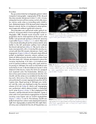
Video 4. TEE 2 and 3D views of the aortic valve showing the left coronary cusp prolapse. View supplemental video at https://doi. org/10.12945/j.jshd.2018.042.17.vid.04.
Video 5. Baseline aortic root angiogram demonstrating the severely regurgitant aortic valve and that the re-implanted coronary arteries were at a safe height from the aortic annu- lus. View supplemental video at https://doi.org/10.12945/j. jshd.2018.042.17.vid.05.
Video 6. Deployment of a 26-mm Sapien-3 valve with aortic root angiogram prior to deployment and rapid pacing during deploy- ment. View supplemental video at https://doi.org/10.12945/j. jshd.2018.042.17.vid.06.
Ghobrial J. et al.
TAVR in TGA
