
Page 26 - Journal of Structural Heart Disease Volume 4, Issue 2
P. 26
47 Case Report
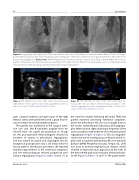
Figure 4. Angiography and angioplasty of the transcatheter aortic valve 10 months post-initial procedure. Panel A. Aortic root angio- gram in the lateral projection demonstrating downward migration of the transcatheter valve of approximately 4–5 mm with severe perivalvular regurgitation (black arrow). Panel B. Angioplasty of the transcatheter Sapien 3 valve using a 24-mm VIDA balloon resolv- ing the residual waist. Note the transvenous pacemaker in the right ventricle. Panel C. Post-angioplasty aortic root angiogram in the lateral projection demonstrating only mild residual perivalvular regurgitation.
Video 9. TEE 2D short axis view of the aortic valve status post deployment of the Sapien-3 valve. View supplemental video at https://doi.org/10.12945/j.jshd.2018.042.17.vid.09.
tures. Surgical cutdown and open repair of the right femoral artery were performed with a good nal re- sult and return of normal peripheral pulses.
The patient was monitored in the surgical inten- sive care unit. Her B-natriuretic peptide level de- creased from 121 pg/ml pre-procedure to 39 pg/ ml. Her post-operative echocardiogram showed no evidence of central or perivalvular regurgitation. She was started on aspirin and clopidogrel and dis- charged on postoperative day 2. On initial clinic fol- low-up within 1 month post-procedure, she reported minimal improvement in her exertional symptoms, and her echocardiogram showed progressive peri- valvular regurgitation (Figure 3; Videos 10 and 11). In
Video 10. TTE 2D long axis view of the aortic valve upon fol- low-up after the procedure demonstrating progressive peri- valvular regurgitation. View supplemental video at https://doi. org/10.12945/j.jshd.2018.042.17.vid.10.
the next few months following the initial TAVR, the patient reported worsening exertional symptoms. Given the perivalvular AR, she was brought back to the cardiac catheterization laboratory. Cineangiogra- phy demonstrated slight downward migration of the aortic prosthesis with evidence of severe perivalvular regurgitation (Figure 4; Video 12). This was hypothe- sizedtobeduetoinadequatepost-dilationoftheSa- pien valve in a patient with primary AR. A 24-mm Vida balloon (BARD Peripheral Vascular, Tempe, AZ, USA) was used to perform high-pressure dilation, which resulted in improved valve apposition to the walls of the hemashield graft and resolution of the perivalvu- lar AR (Figure 4; Videos 13 and 14). The patient toler-
Ghobrial J. et al.
TAVR in TGA
