
Page 25 - Journal of Structural Heart Disease Volume 4, Issue 2
P. 25
Case Report 46
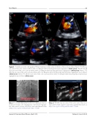
Figure 3. Transthoracic echocardiography (TTE) post-initial procedure and post-dilation of the aortic prosthesis. Panel A. TTE 2D long axis view of the aortic valve prosthesis with color Doppler demonstrating perivalvular regurgitation (white arrow). Panel B. TTE 2D short axis view of the aortic valve prosthesis with color Doppler demonstrating perivalvular regurgitation (white arrow). Panel C. TTE 2D long axis view of the aortic valve prosthesis with color Doppler demonstrating reduced perivalvular regurgitation post dilation (white arrow). Panel D. TTE 2D short axis view of the aortic valve prosthesis with color Doppler demonstrating reduced perivalvular regurgitation post dilation (white arrow).
Video 7. Post-valve deployment aortic root angiogram demon- strating no residual aortic regurgitation or perivalvular regurgita- tion and normal coronary artery ow. View supplemental video at https://doi.org/10.12945/j.jshd.2018.042.17.vid.07.
Video 8. TEE 2D long axis view of the aortic valve status post deployment of the Sapien-3 valve. View supplemental video at https://doi.org/10.12945/j.jshd.2018.042.17.vid.08.
Journal of Structural Heart Disease, April 2018
Volume 4, Issue 2:42-49
