
Page 23 - Journal of Structural Heart Disease Volume 4, Issue 2
P. 23
Case Report 44
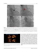
Figure 2. Angiography of transcatheter aortic valve replacement in D-TGA status post arterial switch with a valve sparing root repair using a hemashield graft and repair of severe aortic regurgitation with a 26-mm Edwards Sapien-3 valve. Panel A. Aortic root angiogram demonstrating the severely regurgitant valve with opaci cation of the left ventricle and re-implanted coronary arteries at a safe height from the aortic valve annulus (black arrow marks the aortic valve lea ets). Panel B. The aortic valve in an open position (red arrow). Panel C. Balloon sizing of the aortic valve annulus and aortic valve angioplasty with a waist measuring 19–21 mm. Panel D. Aortic root angiogram status post 26-mm Edwards Sapien-3 valve deployment with no residual regurgitation, no perivalvular regurgitation, and normal coronary artery ow.
Video 3. TEE 3D views of the aortic valve demonstrating the left coronary cusp prolapse; views also used for annular dimen- sions. View supplemental video at https://doi.org/10.12945/j. jshd.2018.042.17.vid.03.
al and ventricular septal defect closure at 2 years of age; and valve-sparing aortic root replacement with a 22-mm Hemashield graft and subaortic membrane resection at 15 years of age. The last operation was complicated by cognitive injury and memory loss, and her recovery was protracted. She subsequently developed symptomatic severe AR. Because of her refusal to receive blood transfusions due to her Jeho- vah’s Witness faith and history of three surgeries, two median sternotomies, and post-operative cognitive injury, she was considered high risk for repeat surgi- cal intervention and was referred for o -label use of TAVR.
Journal of Structural Heart Disease, April 2018
Volume 4, Issue 2:42-49
