
Page 21 - Journal of Structural Heart Disease Volume 4, Issue 5
P. 21
Original Scientific Article 216
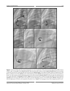
Figure 1. Selective Left Main Coronary Artery Angiograms in a 4 yr. young female child with left anterior descending (LAD) coronary artery to right ventricle (RV) fistula. Panel A. Dilated LAD terminating with a fistula to the RV (arrow). Panel B. Diagnostic JR catheter in left main and an 0.016”Transend guide wire all the way to RV (arrow). Panel C. A 4Fr. Balloon tipped catheter was advanced in LAD. Balloon inflated to block flow (arrow) and this delineated fistula better. Panel D. Cine fluoroscopy of the Excelsior Microcatheter in fistula. Two radio-opaque markers delineating position of the Microcatheter (arrows). Panel E. Cine fluoroscopy after deployment of first Target detachable coil (arrow)[8mmx20cm]. Panel F. Cine fluoroscopy after 4 additional Target detachable coils were deployed (arrow)[second coil was also 8mmx20cm, 3rd and 4th coils were 4mmx8cm and last coil was 6mmx20cm]. Panel G. Angiogram just after the deployment of the five coils revealed no flow distal to coil. Panel H. Final angiogram shows flow stopped proximal to coils.
Journal of Structural Heart Disease, October 2018 Volume 4, Issue 5:212-221
