
Page 22 - Journal of Structural Heart Disease Volume 4, Issue 5
P. 22
217
Original Scientific Article
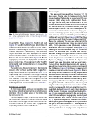
Video 1. Video which illustrates the case mentioned in fig- ure 1. View supplemental video at https://doi.org/10.12945/j. jshd.2018.043.17.vid.01.
tal part of the fistula (Figure 1D). The first coil used (Figure 1E) was 8mmx20cm Target detachable coil (Manufactured by Boston Scientific for Stryker Neuro- vascular). The advantage of such coils is its electrically released mechanism. Subsequently, four additional Target detachable coils were deployed (the second coil was also 8mmx20cm, 3rd and 4th coils were 4mmx- 8cm and last coil was 6mmx20cm (Figure 1F). Repeat angiography between coil deployment was done to assess residual flow. Final angiogram after the fifth coil revealed good coils position and no residual flow (Figure 1G, H).
The patient was allowed to recover in the intensive care unit. After six hours and due to the sluggish cor- onary flow at the end of the procedure (Figure 1G, H), heparin drip was initiated at 15 units/kg/hr keeping PTT at 1.5 times normal. The same evening, the pa- tient received 75mg aspirin and 2mg Warfarin. She was discharged home after 2 days on 75mg aspirin and 2 mg Warfarin. For video of the case, see Video 1.
Anterograde Approach
As mentioned above, a closure can be done from
the venous side either by creating an arteriovenous wire loop [16] or via direct access of the fistula from the venous side [22].
The wire loop technique: to do so, one has to cross the fistula from the arterial side and advance a wire until it exits into the right side and then snare and ex- teriorize from either the femoral vein or jugular vein depending on the location of the fistula.
Case example:
We previously have published this case [17]. 12 days young female baby, 2.4 kg presented in florid congestive heart failure due to a very large left main coronary (LMC) artery to the right ventricle fistula. Her right femoral artery was occluded due to a prior cardiac catheterization. Access was achieved from the left femoral artery 4Fr, left femoral vein 4Fr, and right internal jugular vein 8Fr. The initial hemodynamic as- sessment revealed systemic pulmonary artery pres- sure and infinite Qp:Qs ratio. Angiography in the left main coronary artery revealed the presence of huge LAD to right ventricle fistula (Figure 2A, B). The fistula was crossed easily from the arterial side using a 0.035” floppy tip guidewire. The wire was advanced all the way to the main pulmonary artery and snared using a 4Fr., 10mm gooseneck snare (Microvena) and was exteriorized from the right jugular vein (Figure 2C, D) creating an arteriovenous wire loop. An 8Fr. Mullins sheath was advanced over this wire from the jug- ular vein through the right ventricle into the fistula and into the distal LAD. The first device used was a 12mm Amplatzer muscular VSD device (AGA Medical, Plymouth, MN)[Figure 2E). A total of 7 Flipper coils (five of them were 5mmx8cm, and two were 5mmx- 10cmm (Cook Medical, Bloomington, IN) were de- ployed from the arterial side to create a nest behind the VSD device (Figure 2F, G). Repeat angiogram still revealed significant residual shunt (Figure 2H). Due to the heavy contrast load used (7ml/kg), the procedure was terminated. The baby remained stable without a rise in troponin or lactate but remained intubated with the continued moderate residual flow by echo- cardiography. Therefore, two days later the baby was brought back to the catheterization laboratory and a right carotid artery cut down was used and an 8Fr. sheath was inserted. A 10/8 mm Amplatzer Duct Oc- clud (AGA Medical)(Figure 2I) and a 9mm Gianturco Grifka Vascular Occlusion Device (Cook Medical)(Fig- ure 2J) were deployed proximal to the coils and mus- cular VSD device. Repeat angiography revealed good devices positions and minimal residual flow (Figure 2K, L). Repeat hemodynamics revealed that the pul- monary artery pressure dropped to 40% systemic and the Qp:Qs ratio decreased to 2.7:1. The baby was ex- tubated two days later. She was transferred back to referring institution on 4mg/kg aspirin orally. She was
Al-Qahtani A. et al.
Catheter Closure of Coronary Fistulae
