
Page 24 - Journal of Structural Heart Disease Volume 4, Issue 5
P. 24
219 Original Scientific Article
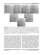
Figure 3. Panel A. Selective left main coronary angiogram in a 76 yr. old gentleman showing very narrow and tortuous circumflex (medium arrow). There is a fistula arising from circumflex (long arrow) and this fistula drained to the right pulmonary artery (short ar- row). Panel B. Selective right coronary angiogram showing the posteromedial branch draining into a fistula (short arrow) that drains into the fistula that arose from the circumflex, all drain into the right pulmonary artery (long arrow). Panel C. Angiogram in LMC artery where fistula drains into mouth of right pulmonary artery, where a 5Fr. JR catheter was positioned (arrow). Panel D. Cine fluoroscopy during deployment of 12mmx30cm Target detachable coil (arrow) using the Excelsior Microcatheter. Panel E. angiogram after de- ployment of additional 7 Target detachable coils (2nd coil: 10mmx30cm; 3rd and 4th coils: 9mmx20cm; 5th, 6th and 7th coils: 4mmx8cm and 8th coil: 3mmx4cm) showing filling of the circumflex (arrow). Panel F. few frames later showing contrast up to the coils (arrow) and (Panel G), few frames later no residual flow in fistula and good coils position (arrow).
phy in the left main coronary artery revealed normal LMC and LAD. The circumflex origin was very narrow and very tortuous. At mid circumflex, a fistula arose and drained to the right pulmonary artery (RPA). The fistula size was double the size of the circumflex (Fig- ure 3A). Angiography in the right coronary artery re- vealed normal artery with minimal coronary artery disease; however, the distal branch (posteromedial coronary artery) drained via smaller channels and connected with the circumflex fistula and all drained to the RPA (Figure 3B). Multiple attempts to cross the fistula from the LMC artery failed. Therefore, a 5Fr. JR catheter was used from the venous side to the RPA and crossed the exit site of the fistula (Figure 3C). A
0.016”guidewire (Transend) was used to navigate the fistula. The wire and a 150cmx6cm Excelsior SL-10 Mi- crocatheter were advanced all the way to the origin of the fistula from the circumflex. The wire was removed and a total of eight Target detachable coils were de- ployed in mid-distal fistula (1st coil: 12mmx30cm; 2nd coil: 10mmx30cm; 3rd and 4th coils: 9mmx20cm; 5th, 6th and 7th coils: 4mmx8cm and 8th coil: 3mmx4cm (Fig- ure 3D, E, F). Repeat angiography revealed complete closure of fistula (Figure 3G). The patient had recov- ered overnight and a repeat echocardiography the next day revealed complete closure of the fistula. He was discharged home after 24 hours from the proce- dure on his medications of warfarin and aspirin.
Al-Qahtani A. et al.
Catheter Closure of Coronary Fistulae
