
Page 23 - Journal of Structural Heart Disease Volume 4, Issue 5
P. 23
Original Scientific Article 218
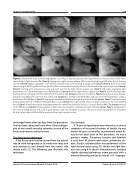
Figure 2. Selective left main coronary angiograms in a 2.4 kg 12 days young baby with large left main coronary artery (LMC) fistula terminating in right ventricle (RV). Panel A. Angiogram in right anterior oblique (RAO) view showing large LMC artery fistula draining into RV. Panel B. Repeat angiogram in left anterior oblique (LAO) view showing drainage to RV (arrow). Panel C. Cine fluoroscopy after passing an 0.035” guide wire from fistula to RV to main pulmonary artery where it was snared using a gooseneck snare (arrow). Panel D. Forming of an arteriovenous wire loop with exit into the right internal jugular vein. Panel E. LMC artery angiogram after deployment of a 12mm Amplatzer muscular VSD device (arrow) from the right internal jugular vein. Panel F. Cine fluoroscopy after deployment of 1st Flipper coil (5mmx8cm) from the arterial side (arrow) proximal to the device. Panel G. Cine fluoroscopy after de- ployment of additional 6 coils from the arterial side (arrow)[four of them were 5mmx8cm and two were 5mmx10cmm] to create a nest behind the VSD device. Panel H. Angiogram revealed residual flow through the fistula to the RV (arrow). Panel I. Cine fluoroscopy during deployment of a 10/8mm Amplatzer Duct Occlud (arrow) from right carotid artery cut-down proximal to the coils and VSD device. Panel J. Cine fluoroscopy during deployment of a 9mm Gianturco Grifka Vascular Occlusion Device (GGVOD) (arrow) proximal to the PDA device. Panel K. Final angiogram in RAO view showing near complete occlusion of fistula (arrow). Panel L. Angiogram in LAO view showing devices with coils in good position with near complete occlusion. Thin arrow shows opacification of circumflex, fat short arrow shows LAD and fat long arrow shows right coronary artery.
discharged home after ten days from the procedure. She had been doing well since then. Echocardiogra- phy at one month revealed complete closure of the fistula and normal cardiac function.
The direct access technique
If the fistula could not be crossed from the arterial
side to close retrogradely or to create wire loop, one may attempt to cross directly from the venous side (fistula exit) [22]. The following case illustrates this technique.
Case Example:
A 76-year-old gentleman was referred to us due to symptoms of increased shortness of breath. He was known to have a circumflex to pulmonary artery fis- tula for ten years prior to this procedure. He was a previous smoker. Pulmonary function test revealed a mild form of chronic obstructive pulmonary dis- ease. Cardiac catheterization was performed via the right femoral artery using 7Fr. sheath and right fem- oral vein using 6 Fr. sheath. Hemodynamics revealed slightly elevated pulmonary artery pressure with a mean of 26mmHg and Qp:Qs ratio of 1.4:1. Angiogra-
Journal of Structural Heart Disease, October 2018
Volume 4, Issue 5:212-221
