
Page 46 - Journal of Structural Heart Disease Volume 4, Issue 5
P. 46
241 Case Report
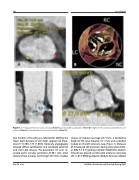
Figure 1. CT measurements of aortic annulus (Panel A), aortic leaflet calcification (Panel B), height of left coronary ostia/left sinus of Valsalva (Panel C) and diameters of sinuses of Valsalva (Panel D).
tion fraction (72%) who was referred for TAVR by the Heart Team because of very high surgical risk (Euro- score ll 15.30%, STS 11.06%). Coronary angiography showed diffuse calcification and moderate proximal and mid LAD disease. Pre-procedural CT scan re- vealed aortic annulus perimeter of 60.1 mm, short distance from annulus to LM origin (6.5 mm), shallow
sinuses of Valsalva (average 26.7 mm), a borderline height of left sinus Valsalva (15.1 mm) and a calcified nodule on the left coronary cusp (Figure 1). Because of threatened LM occlusion during valve placement, an EBU 3.5 6 Fr guiding catheter (Medtronic, Dublin, Ireland) was placed via left radial artery to cannulate LM. A .014” BMW guidewire (Abbott Vascular, Abbott
Noc M. et al.
Left Main Protection and Stenting During TAVR
