
Page 47 - Journal of Structural Heart Disease Volume 4, Issue 5
P. 47
Case Report
242
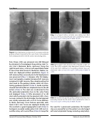
Figure 2. Valve deployment to the point of no recapture with the guiding catheter lying behind the valve frame, and guidewire with undeployed stent in the left anterior descending artery.
Park, Illinois, USA) was advanced into LAD followed by placement of undeployed drug-eluting stent Or- siro 3.5x15 (Biotronik, Berlin, Germany). Using the right femoral artery, a 26 mm self-expandable Evolut R valve (Core Valve Evolut R, Medtronic, Dublin, Ire- land) was deployed without predilation (Figure 2). Left coronary flow, assessed prior to full deployment, was preserved (Video 1). However, after full deploy- ment, aortography revealed decreased left coronary compared to right coronary flow despite pulling the stent back to the guiding catheter while still main- taining guidewire position (Video 2). Guide injection showed that left leaflet was displaced close to the LM ostium (Video 3). The stent was re-advanced to the LM, protruded proximally close to the valve frame and deployed (Video 4). After placement of an LM stent which extended into the proximal LAD, a hazi- ness was noticed at the distal edge. Stented segment was initially extended with a 2.5x12 Orsiro (Biotron- ik, Berlin, Germany). Since haziness persisted, addi- tional 2.5x15 mm Orsiro was deployed distally and this resulted in a good angiographic result (Video 5). Aortogram revealed widely patent LM, LAD and cir- cumflex artery with normal flow (Video 6). Except for
Video 1. Aortogram before complete valve deployment. Both coronary arteries are well perfused. View supplemental video at https://doi.org/10.12945/j.jshd.2018.008.18.vid.01.
Video 2. Aortogram with decreased left compared to right cor- onary flow after complete valve deployment despite moving of the stent from LAD back to the guiding catheter. View sup- plemental video at https://doi.org/10.12945/j.jshd.2018.008.18. vid.02.
Video 3. Injection through the guiding catheter revealed a mass protruding toward the LM ostium. View supplemental video at https://doi.org/10.12945/j.jshd.2018.008.18.vid.03.
the need for a permanent pacemaker, the hospital stay was uneventful and the patient was discharged with dual antiplatelet therapy including acetylsalicyl-
Journal of Structural Heart Disease, October 2018
Volume 4, Issue 5:240-245
