
Page 48 - Journal of Structural Heart Disease Volume 4, Issue 5
P. 48
243
Case Report
Video 4. Stent deployment from the LM toward the valve frame. View supplemental video at https://doi.org/10.12945/j. jshd.2018.008.18.vid.04.
Video 5. Post procedural guide injection showed widely patent LM, LAD and left circumflex artery with normal flow. View sup- plemental video at https://doi.org/10.12945/j.jshd.2018.008.18. vid.05.
Video 6. Final aortogram with comparable left and right coro- nary flow. View supplemental video at https://doi.org/10.12945/j. jshd.2018.008.18.vid.06.
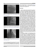
ic acid and clopidogrel. Post-procedural CT scan after three weeks revealed widely patent LM stent protrud- ing across the sinus of Valsalva close to valve frame
with its proximal part in a “T-stent” shape. (Figure 3). Twelve months after the intervention, the patient continues to be asymptomatic.
Discussion
Several anatomic factors derived from preproce- dural CT scan including low LM ostium, shallow sinus- es of Valsalva, severe leaflet calcification with large bulky calcium nodules, high native leaflet length/ curved coronary sinus height ratio as well as extreme valve oversizing and “valve-in-valve” procedure, have been identified as high risk features for coronary oc- clusion during TAVR [2, 4]. A potential risk of coronary occlusion may also be assessed before TAVR using aortic valve predilatation with simultaneous aorto- gram. If the coronary occlusion is documented, up- front LM protection is mandatory [4]. We did not use this technique because we perform a vast majority of TAVR without predilatation. If we predilate, we always use a small balloon (18-20 mm) to minimize manipu- lation of the calcified native valve. Predilatation with a smaller balloon would probably underestimate the likelihood of actual LM occlusion during TAVR. More- over, based on the CT scan, we have already decided to use upfront LM protection. Admittedly, according to CT-derived measurements, a 23 mm rather than 26 mm Evolut R should have been used. Some oversiz- ing with this self-expandable prosthesis was select- ed because annulus perimeter was at the very upper limit for the 23 mm valve, the native valve was highly calcified, and we have already decided for upfront LM protection.
The optimal strategy for LM protection during TAVR, particularly when using self-expandable Evo- lut R valve, remains to be defined. The "chimney" technique is generally considered in patients with low sinotubular junction due to potential occlusion of the respective sinus of Valsalva after valve deploy- ment. In our patient, the height of the left sinus of Val- salva was just above the recommended 15 mm. We, therefore, decided for a less complex “T-stenting” and protrude LM stent only above the displaced leaflet and toward the valve frame. Accordingly, the proxi- mal part of the stent was not behind the valve frame. This avoids possible unfavorable interaction between the LM stent and self-expanding valve frame which
Noc M. et al.
Left Main Protection and Stenting During TAVR
