
Page 29 - Journal of Structural Heart Disease Volume 5, Issue 1
P. 29
Case Report
18
output. This could be attributed to the fact that the existed from the time of surgery residual infundibu- lar stenosis had not yet completely regressed to per- mit the maintenance of a sufficient pulmonary blood flow exclusively though the native RNOT. After discus- sion with our surgeons and the family, in 2008, an 18 mm Melody® valve was placed successfully in the Go- re-Tex tube. After the valve implantation the patient improved and he was doing quite well but 6 years later he developed again progressively aggravated exercise intolerance (NYHA class II –III symptoms) due to chronic pulmonary regurgitation this time through its operated native RVOT. In December 2017 echo- cardiographic and CMR evaluation revealed signifi- cant pulmonary regurgitation through the surgically placed transannular patch resulting in progressive dilatation of the RVOT and the pulmonary arteries. Doppler echocardiographic study showed a normal functioning Melody valve with no residual gradient and regurgitation. The CMR estimated RVEDVi and RVEF were 165 ml/m2 and 48%, respectively. After the CMR study, the patient underwent cardiac cath- eterization with complete hemodynamic and an- giographic studies. There was a 30 mm Hg pressure gradient across the RVOT. The maximum systolic di- ameters of the proximal RVOT (just below the valve level), estimated valve annulus, proximal and distal MPA measured using biplane angiography, were 31 mm, 26 mm, 32mm and 34. 5 mm, respectively. The length from the RVOT from the estimated valve annu- lus position to the PA bifurcation was 35 mm. Balloon sizing of the pulmonary arteries was performed with maximal inflation of an ASD 34 mm Amplatzer sizing balloon simultaneously with an ascending aortogram that excluded coronary artery proximity and obstruc- tion. Balloon sized RVOT/MPA diameters were only 1 mm larger than the angiographic ones. After obtain- ing procedural permission on compassionate basis from the Greek Federal Drug Administration and a written consent from the parents and the patient, it was decided to proceed to percutaneous pulmonary valve implantation with Venus P-valve.
To ensure stability and good opposition to the wall of the MPA, the Venus P-valve was selected to be 4 mm larger than the measured balloon waist at the pulmonary valve annulus level and equal to the maximal angiographic systolic diameter of the MPA.
Video 1. Injection of contrast medium through a pig-tail 5F cath- eter into the proximal LPA showing competent Venus P-valve and Melody valves with no pulmonary regurgitation. View sup- plemental video at https://doi.org/10.12945/j.jshd.2019.024.18. sup.01.
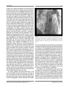
The valve implantation was performed under general anesthesia using the previously described technique. [2-4] Based on the angiographic measurements and the anatomic characteristics of the RVOT, a 36 mm di- ameter and 35 mm length flared Venus P-valve was successfully implanted from the RVOT to MPA bifurca- tion through a 24 F delivery system. (Figure 1 A,B,C,D). The procedure was guided using frequent check an- giograms through a Berman angiographic catheter placed in the MPA/RV from a second femoral venous approach. Post procedural pulmonary angiography and hemodynamic measurements showed a well positioned and opposed to the wall of MPA compe- tent valve and no pressure gradient across the RVOT (Video 1). The procedure and radiation times were 312 minutes and 48 minutes, respectively.
The patient was discharged the next day of the procedure on oral aspirin 3-5 mg/Kg/day for life. Be- fore discharge an electrocardiogram (ECG) recording, a biplane chest X-ray, and a transthoracic echocardi- ography (TTE) were performed. Fluoroscopic exam- ination 3 and six months after the procedure showed a good valve position with no fractures (Figure 3). At
Journal of Structural Heart Disease, February 2019
Volume 5, Issue 1:16-20
