
Page 30 - Journal of Structural Heart Disease Volume 5, Issue 1
P. 30
19 Case Report
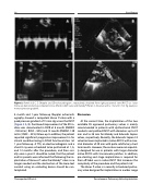
Figure 2. Panels A, B, C, D. Doppler and 2D echocardiogram , respectively, obtained from right parasternal view (RVOT) at 1-year follow-up demonstrating competent Venus (Panels A, B) P-valve and Melody® (Panels C, D) valves with a 15 and 21 mm Hg peak pul- monary pressure gradient.
6 month and 1-year follow-up Doppler echocardi- ography showed a competent Venus P-valve with a peak pressure gradient of 15 mm Hg across the RVOT (Figure 2 A, B). Continued improvement of the RV in- dices was documented in CMR at 6 month (RVEDVi -150 ml/m2, RVEF – 50%) and 12 month (RVEDVi -120 ml/m2, RVEF – 55%) follow-up. In addition, the patient reported significant progressive improvement in his clinical condition being in NYHA functional class I at 1 year follow-up. A TTE, an electrocardiogram and a chest-X ray were scheduled to be performed at 1, 6, and 12 months after the procedure, and then seri- ally once a year. It should be noted, that the patient and his parents were informed that following the im- plantation of Venous-P valve the Melody® valve is no longer needed and the obstruction of the Gore-text conduit using an occluding device should be con- templαted.
Discussion
At the current time, the implantation of the two available CE approved pulmonary valves is mainly recommended in patients with dysfunctional RVOT conduits and patched RVOT with diameters up to 22 mm and to 26 mm for Melody and Edwards Sapien valves, respectively. Recently, the Edwards Sapien S3 valve has been implanted in native RVOTs with a max- imal diameter of 29 mm with quite satisfactory short term results. However, these valves were not especial- ly designed for use in patients with larger diameter native RVOTs with transannular patches. In addition, pre-stenting and stage implantation is required for their off-label use in native RVOT that increases the complexity of the procedure and the patient risk.1
The Venus P-valve is a recently introduced pulmo- nary valve designed for implantation in a wider range
Thanopoulos B.D. et al.
Percutaneous Pulmonary Valve Implantation
