
Page 15 - Journal of Structural Heart Disease Volume 5, Issue 3
P. 15
Original Scientific Article
54
100–280 mAs per rotation for CCT. A non-enhanced electrocardiography (ECG)-gated CT scan prospec- tively triggered at 75% of the R-R interval was per- formed to measure the aortic valve calcium score. For CCT, ECG-based tube current modulation was not implemented. Contrast agent application was controlled by a bolus tracking technique using 80 to 120 ml of contrast media (Isovue-370, Iopamidol In- jection 76%, Bracco Diagnostics, Inc). Ten transaxial data sets were reconstructed with retrospective ECG gating at 10% steps from 0–90% of the R-R interval for each patient.
CCT image analysis
All data were transferred to a dedicated worksta- tion (Syngo Via software, Siemens Medical Solutions, Forchheim, Germany). Analysis of CCT images was performed by a cardiac radiologist (13 years of expe- rience with CCT) who was blinded to patient clinical data including all clinical findings, history, and TTE results. Repeat assessments were performed by the
same radiologist at least 1 month apart in random or- der to prevent recall bias.
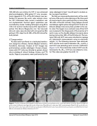
The AVA was measured by planimetry of the small- est area of the aortic valve opening on the time point of maximal aortic valve opening (early or mid-systole, 10%-20% of the R-R interval), using oblique coronal and oblique sagittal planes along the LVOT and an ad- ditional oblique transverse plane parallel to the aor- tic valve. The largest cross-sectional area of the LVOT was measured at the hinge point of the insertion of 3 aortic cusps on the double-oblique transverse plane during mid-systole (20% of the R-R interval). The ana- tomic AVA and LVOT area were calculated as average of 2 planimetric measurements using an electronic caliper [13]. The following measures were obtained: LVOT minimal and maximal diameters (Dmin and Dmax) and LVOT area excluding aortic annulus calcification (Figure 1) [17]. The eccentricity index of LVOT was de- termined as Dmin/Dmax. LVOT was considered as circu- lar if the index was greater than 0.9 [13].
Figure 1. Cardiac computed tomography image analysis before transcatheter aortic valve replacement includes (Panel A) degree of aortic valve calcification and Agatston aortic valve score, (Panel B) aortic valve area (AVA) measurement, (Panel C) left ventricular out- flow tract (LVOT ) area measurement without calcification, (Panel D) LVOT area measurement with calcification, and (Panel E) minimum (Dmin) and maximum (Dmax) diameters of LVOT.
Journal of Structural Heart Disease, June 2019 Volume 5, Issue 3:52-61
