
Page 30 - Journal of Structural Heart Disease Volume 5, Issue 4
P. 30
Meeting Abstracts
92
30. Figure 4. The procedure was done, performing A-V loop through a 4 Fr. Torq Vue catheter (NR), releasing one ADO II 6/4 device from the right side of the CAF.
X ray show normal pulmonary blood flow and a better car- diothoracic ratio (Figure 4).
Echocardiogram demonstrated a tiny residual shunt, smaller cardiac chambers and no signs of pulmonary hypertension (Figure 5).
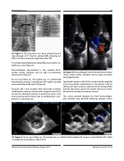
At one year follow up the patient was re- catheterized founding left coronary trunk dilated, CAF totally occluded and normal flow in DA and CX (Figure 6).
Clinically CAF could manifest with myocardial ischemia resulting from coronary steal or with congestive heart fail- ure due to substantial systemic-to pulmonary shunt. Such early postnatal presentation, due to hemodynamic com- promise is extremely rare.
30. Figure 5. Echocardiogram demonstrated a tiny residual shunt, smaller cardiac chambers and no signs of pulmo- nary hypertension.
Symtomatic patients with CAF must be treated surgically or by transcatheter embolization. In the present case the absence of major coronary collateral vessels arising within the CAF, the unique course of it and the absence of multi- ple drains favoured percutaneous closure.
The correct prenatal diagnosis by fetal echocardiogra- phy, enabled close perinatal follow-up, prompt clinical
30. Figure 6. At one year follow up the patient was re- catheterized founding left coronary trunk dilated, CAF totally occluded and normal flow in DA and CX.
Journal of Structural Heart Disease, August 2019 Volume 5, Issue 4:75-205
