
Page 31 - Journal of Structural Heart Disease Volume 5, Issue 4
P. 31
93
Meeting Abstracts
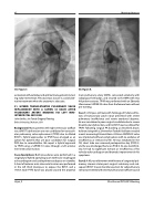
30. Figure 7.
evaluation without delay and optimal management, includ- ing early intervention. Percutaneous closure is a valid alter- native treatment when the anatomy is adecuate.
31. HYBRID TRANSCATHETER PULMONARY VALVE REPLACEMENT WITH A SAPIEN S3 VALVE AFTER PULMONARY ARTERY BANDING VIA LEFT MINI- INTERCOSTAL INCISION
John Serfas, Joe Turek, Gregory Fleming
Duke University, Durham, USA
Background: Many patients with right ventricular outflow tract (RVOT) dysfunction are not candidates for transcath- eter pulmonary valve replacement (TPVR) due to dilated RVOT’s. Hybrid approaches to TPVR have emerged as an option for patients that are poor candidates for surgical PVR due to comorbidities. We report a hybrid approach to TPVR using a SAPIEN S3 valve through a left anterior mini-intercostal incision.
Case descriptions: Both procedures were performed in a single plane hybrid operating room with trans-esophageal echocardiogram and cardiopulmonary bypass on standby. A 5cm left anterior mini-intercostal incision was performed in the left third interspace to expose the RVOT, and a 14mm wide PTFE band was placed around the proximal
30. Figure 8.
main pulmonary artery (MPA), connected anteriorly with radiopaque hemoclips, and secured to the MPA with two 4-0 prolene sutures. TPVR was performed with an Edwards Lifesciences SAPIEN S3 valve from the femoral vein without pre-stenting.
Case 1: A 58 year old male with Tetralogy of Fallot and his- tory of transannular patch repair presented with severe pulmonary insufficiency and severe exertional dyspnea. He was considered a poor surgical candidate due to severe biventricular dysfunction, and his RVOT was too dilated for TPVR. Following surgical PA banding as described above, balloon sizing with a 25mmx4cm Tyshak II balloon revealed a waist measuring 22mmx24mm. A 26mm SAPIEN S3 valve was implanted without complications with no evidence of insufficiency or stenosis by TEE. He was extubated in the OR, chest tube was removed postoperative day (POD) 2, and he was discharged home on POD 4. At one month fol- low, he had no significant stenosis or insufficiency of his valve by echocardiogram and dramatic improvement in his symptoms.
Case 2: A 65 year old women with history of congenital pul- monary stenosis status-post surgical valvotomy and sub- sequent RVOT muscle bundle resection for subpulmonary stenosis presented with severe pulmonary insufficiency and
Hijazi, Z
22nd Annual PICS/AICS Meeting
