
Page 25 - Journal of Structural Heart Disease Volume 5, Issue 5
P. 25
Original Scientific Article 218
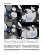
Figure 3. Computerized tomography of a 70-year-old patient in sinus rhythm. Panels A and B show multiplanar reconstructions of the left atrial appendage (LAA) at 45% of the peak R-wave to R-wave. Panel A. Tracing of the LAA ostium showing an area of 446 mm2. Panel B. plane of the LAA ostium. The arrows mark the left circumflex coronary artery. Panels C and D show multiplanar reconstruc- tions of the same patient at 5% of the peak R-wave to R-wave with significantly smaller LAA dimensions. Panel C. Tracing of the LAA ostium showing an area of 188 mm2. Panel D. plane of the LAA ostium.
ventricular diastolic phases for both sinus rhythm and atrial fibrillation patients (75% of the peak R-wave to R-wave). Similar to our observation – albeit with com-
parisons only in ventricular diastolic phases- the au- thors report significantly larger dimensions of LAA-or- ifice as well as LAA volume in AF patients compared
Journal of Structural Heart Disease, October 2019
Volume 5, Issue 5:213-220
