
Page 14 - Journal of Structural Heart Disease Volume 5, Issue 6
P. 14
Original Scientific Article
238
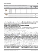
Table 1. Different types of MFO catalog numbers with the corresponding delivery sheaths.
Catalog Number
D Disc Diameter (mm)
D2 Waist Diameter LV Side (mm)
D1 Waist Diameter RV Side (mm)
L Waist Length (mm)
Recommended Delivery Sheath (Fr)
LT-MFO-5-3
LT-MFO-6-4
LT-MFO-7-5 LT-MFO-8-6
LT-MFO-9-7
LT-MFO-10-8
LT-MFO-12-10
LT-MFO-14-12
10 5
10 6
12 7 12 8
14 9
14 10
16 12
18 14
3
4
5 6
7
8
10
12
4
4
4 4
4
4
4
4
4F-5F
6F 7F
Although surgical intervention is considered the gold standard for VSD [3-6], VSDs can successfully be closed by catheterization in patients with favorable anatomies and precise indications. Despite this, dif- ferent complications have arisen where the complete AV block [7, 8], remains the most fearsome, and there- fore, it is necessary to be extremely careful with the indication of this procedure, the choice of the device, and even with the vascular access selection.
Different devices have proven to successfully close different types of VSDs, depending on the size, lo- cation, and presence or absence of prolapse of the aortic sigmoid. Several devices have been used in VSD closure, such as the PDA Nit-Occlud® coil, Flip- per® PDA [9, 10] coil, AMPLATZERTM [11, 12] devices for muscular VSD and Ductus ADO II devices [13, 14].
Regarding vascular access, the arteriovenous loop for the perimembranous VSDs that requires puncture of the femoral artery and the femoral vein was al- ready described by Lock et al [15]. In mid-ventricular and apical VSDs, the arteriovenous loop is performed through the right jugular vein. However, the retro- grade approach from the femoral artery can be done by using devices with symmetrical discs that allow easy access and rapid treatment of the VSD [3, 12].
The new Lifetech KONAR-MFTM Multifunctional Oc- cluder (MFO) was developed to allow the occlusions of small to large defects that can be placed through both vascular approaches, anterograde and retrograde.
The purpose of this study is to report the short- and mid-term results of perimembranous and mus- cular VSDs closure with the use of the new MFO.
Materials and Method
The experience with the new MFO for endovascu- lar closure of VSDs began in October 2017.
Device design
The MFO is a self-expanding occluder consisting of a layer nitinol wire mesh with 144 wires of 0.002” ni- tinol cables. It has two discs joined by a waist, which is formed by a truncated cone. The base of this cone is attached to the left disc and from the vertex, which is umbilicated, hangs an arm that joins it to the right disc. This arm allows articulation to the right disc. The length of the waist is 4 mm and stretches up to 7 mm.
The left disc or “high-pressure disc” is attached to the base of the truncated cone of the waist, and the right or “low-pressure disc” is attached to the waist arm. Each disc contains a 2.4 mm long hub with a screw so that the device can be positioned retro- gradely or anterogradely. Both discs of the device are of equal size. It is a symmetric device.
The base of the cone or “D2” has a diameter of 2 mm greater than the vertex. adding 2 mm on each side to “D2” we obtain the diameter of the left disc or
Journal of Structural Heart Disease, December 2019
Volume 5, Issue 6:237-247
