
Page 16 - Journal of Structural Heart Disease Volume 5, Issue 6
P. 16
Original Scientific Article 240
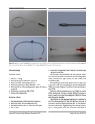
Figure 2. Delivery System. Panel A. The cable has, on one side a screw to hold the device and on the other side, the handle. Panel B. The screw that holds the device. Panel C. The sheath. Panel D. The loader with the device.
Protocol design
Inclusion criteria
1. Patient > 2.5 kg
2. Perimembranous VSD with aneurism
3. Muscular VSDs with adequate rims
4. Pulmonary/Systemic flow (QP/QS) > 1.5:1
5. Clinical and/or echocardiographic signs of volume
overload
6. Pulmonary resistance (PR) ≤7UW
7. History of endocarditis
Exclusion criteria
1. Perimembranous VSDs without aneurysm
2. Muscular VSDs with inadequate rims
3. Not perimembranous or muscular VSD type
4. PR > 7 UW
5. Associate congenital heart disease of exclusively surgical resolution
All previous measurements for the patient selec- tion were made with transthoracic echocardiography (TTE), included the right orifice, the left orifice, and the VSD length.
During the procedure, the patients over 5 Kg, were measured with transesophageal echocardiography (TEE) and under 5kg by transthoracic echocardiogra- phy (TTE).
Patients with perimembranous and high muscular VSDs performed 24 hours Holter before the proce- dure as well as an electrocardiogram (EKG) for those with the prior disorder.
The choice of the appropriate device is related to the TTE measurements of the VSD orifices, the size of the waist and the high-pressure disc of the device. The waist diameter, suggested by the device Compa- ny, is 2 mm greater than the maximum diameter of
Journal of Structural Heart Disease, December 2019
Volume 5, Issue 6:237-247
