
Page 18 - Journal of Structural Heart Disease Volume 5, Issue 6
P. 18
Original Scientific Article 242
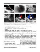
Figure 4. Muscular VSD. Panel A. TEE 0-degree view. The arrow shows the Muscular VSD. Panel B. Left Ventricle Angiography (4 cham- bers): The Muscular VSD is shown by the arrows. Panel C. MFO Anterograde approach positioning. Panel D. Left Ventricle Angiography (4 chambers): The device is correctly in place. Panels E and F. TEE shows no residual shunt.
• Muscular VSD: 8 patients (5 high-muscular and 3 mid-ventricular VSDs). 1 patient had a Ductus Arte- riosus associated that was also closed in the same procedure.
• 2 postoperative VSDs: 1 patient had a Perimem- branous VSD surgically repaired who suffered from endocarditis and presented with a residual shunt, and 1 patient had Tetralogy of Fallot surgically re- paired with a residual VSD and Subtricuspid left ventricle-right atrium shunt.
• 2 postoperative VSDs: 1 patient had a Perimem- branous VSD surgically repaired who suffered from endocarditis and presented with a residual shunt; The other patient had Tetralogy of Fallot surgically repaired with a residual VSD and subtricuspid left ventricle-right atrium shunt.
VSD echocardiographic measurements:
• Left orifice: mean 6.92 mm ±SD 2.83.
• Right orifice: mean 4.54 mm ±SD 1.50. 2 patients
had more than one right orifice.
• Length: mean 4.82 mm ±SD 3.72.
Hemodynamic data:
• QP/QS: mean 1.65/1 (1.3/1 to 2.2/1).
• Pulmonary pressure: mean 24/11 (20/10 to 40/16).
The device most frequently used was 7-5. The de- vices employed were: 5-3: 3 devices, 6-4: 2 devices, 7-5: 4 devices, 8-6: 3 devices, 9-7: 1 device and 10-8 1 device.
The mean of the right waist was 5.07mm ±SD 1.52; the mean of the left waist was 7.07mm ±SD 1.52, and the mean length of the waist was 4 mm.
Endovascular VSD closure was successful in 13 out of 14 patients with trivial to no residual shunt. The failed procedure was on a patient who had a high muscular VSD and persisted with severe resid- ual shunt after the implantation of the device. There was an attempt to close the VSD with a bigger device, but the patient presented a transient AV block during the procedure so the decision was taken to surgically close the defect.
Journal of Structural Heart Disease, December 2019
Volume 5, Issue 6:237-247
