
Page 19 - Journal of Structural Heart Disease Volume 5, Issue 6
P. 19
243 Original Scientific Article
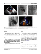
Figure 5. Mid-Muscular VSD. Panel A. TEE: 4 chambers view shows the Mid-ventricular VSD. Panel B. Left Ventricular Angiogram: the arrow shows the VSD Panel C. MFO totally positioned crossing the mitral valve. Panel D. Left Ventricular Angiogram: a trivial residual shunt can be observed. Panel E. TEE: 4 chambers view: the arrow shows the trivial residual shunt.
Follow up
The mean follow-up was 5.75 months (1 to 10 months). No complete AV block, hemolysis or may- or complications were observed throughout the fol- low-up.
< 5 kg
Patients <5 kg had another congenital heart dis- ease associated with severe hemodynamic repercus- sions. All 3 patients presented with heart failure and pulmonary edema.
The mid-ventricular muscular VSDs were the type of defect.
The associated congenital heart diseases were as follow:
Case 1
1-month-old, 3 kg. Suffered from severe Aortic ste- nosis and severe Aortic coarctation associated with patent Ductus Arteriosus and a muscular VSD. Aor- tic valvuloplasty and balloon angioplasty were per- formed and, 4 days later, the Aortic arch was surgical- ly repaired and the Ductus closed. A week later, the patient remained with heart failure and pulmonary edema, so endovascular occlusion of the VSD was successfully done (Figure 5).
Case 2
3-month-old, 3 kg. Presented with transposition of great arteries and a muscular VSD. After being sur- gically repaired with arterial switch technique, the patient persisted with residual VSD and severe heart
Damsky Barbosa J. et al.
VSD Closure with KONAR-MFO
