
Page 20 - Journal of Structural Heart Disease Volume 5, Issue 6
P. 20
Original Scientific Article
244
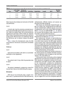
Table 2. Shows the hemodynamic data, VSD measurements, the devices used and the immediate results.
Case
Left To Right Shunt
Pulmonary Hypertension
Left Orifice
Right Orifice
Length
MFO
Residual Shunt
1 Severe
2 Severe
3 Severe
Severe
Severe Moderate
8 MM 5 MM 5 MM
8 MM 7 MM 5 MM
4 MM 10/8 3 MM 8-6 3 MM 6-4
No Mild No
failure reason why endovascular occlusion of the VSD was performed.
Case 3
5-month-old, 5 kg. The anomaly associated was an obstructed infradiaphragmatic pulmonary venous re- turn. After the surgical repair, the severe pulmonary hypertension persisted so Nitric Oxide was required. After 10 days, the pulmonary pressure decreased and endovascular VSD occlusion was performed. During the Pulmonary vein re-stenosis surgery, the patient died.
Table 2 shows the hemodynamic data, the VSD measurements, the devices used and the immediate result.
Follow up
Case 1
Good clinical condition with Aortic re-coarctation waiting for a new angioplasty.
Case 2
The patients died 15 days after the procedure due to sepsis.
Case 3
The patient developed a progressive severe Pul- monary Vein Stenosis and died during the surgery.
Discussion
VSD closure has historically been surgical [3-6]. After Lock [15] performed the first VSD occlusion by
catheterization, different devices and vascular ap- proaches have been proposed.
VSD closure was historically treated through sur- gery [3-6]. After Lock15 performed the first VSD occlu- sion by catheterization, different devices and vascu- lar approaches, such as Nit-Occlud® PDA coil, Flipper® PDA [9, 10] coil and AMPLATZERTM [11, 12] devices for muscular VSD and Ductus ADO II devices [13,14]. have been used to perform the closure.
Few complications were reported, being complete AV block the most feared [7, 8, 17-19].
The endovascular occlusion of perimembranous defects with the MFO device has the same feasibili- ty as other known devices. The MFO great versatility in the vascular access, allow the possibility of closing defects of larger sizes with lower profile sheaths that can be placed through both vascular approaches, an- terograde and retrograde, expanding the spectrum of patients who can benefit from the alternative clo- sure by catheterization, among others.
In the short term follow-up, we did not observe in the cohort patients the complete AV block, however, it is a complication that can appear at any time that requires [19] a close follow-up.
In the vascular access, we usually use the antegrade approach for mid-ventricular and apical muscular de- fects, leaving the retrograde maneuver for the peri- membranous and high muscular VSDs. Depending on the circumstances, avoiding the arteriovenous loop can simplify the procedure and reduce the risks associated with this technique. Moreover, avoiding arterial puncture represents a great advantage, es- pecially in small patients. The MFO’s great versatility, due to the double hub with screw-in both sides, al- lows the occlusion of the VSD in an antegrade or ret- rograde way according to convenience. In complex cases where the placement of the device is challeng- ing, we suggest the use of the antegrade approach.
Journal of Structural Heart Disease, December 2019
Volume 5, Issue 6:237-247
