
Page 45 - Journal of Structural Heart Disease Volume 5, Issue 6
P. 45
269
Case Report
is mostly asymptomatic unless another comorbidity is present. In the literature, there are isolated case re- ports on the absence of the left CCA in combination with other various anomalies of the AA [10-14]. How- ever, there is no case reported in combination with hypoplastic Gothic AA and VSD.
We present the first, to the best of our knowledge, case of staged endovascular treatment for ventricular septal defect (VSD) in combination with AA anoma- ly, represented by an anatomically complex and rare variant of hypoplasia and kinking of the AA (Gothic, elongated above the sternoclavicular joint) along with congenital absence of the left CCA, looping of the right subclavian artery and anomalous origin of the left subclavian artery from the descending tho- racic aorta.
Case presentation
A 10-year-old boy was admitted with complaints of headache, high blood pressure, and fatigue. The patient had a history of congenital heart defect – a VSD and patent ductus arteriosus were diagnosed at birth. At 10 months, he was first diagnosed with the AA anomaly (elongation, hypoplasia, and stenosis due to kinking with SPG of 50 mm Hg), absence of the left CCA, looping of the right subclavian artery in combination with perimembranous VSD of 7.5 mm (Figure 1). At the age of 1 year and 1.5 months, tran- scatheter VSD closure was done using an 8mm Am- platzer Perimembranous Ventricular septal occluder (Figure 2). Four months later (at the age of 1.5 years), transluminal balloon angioplasty for the AA and AI stenosis was performed using a Tyshak II balloon catheter (NuMED Inc., Hopkinton, NY) with a diame- ter of 8 mm, the SPG decreased from 53 to 35 mm Hg. Subsequently, the child was observed on an outpa- tient basis. According to the mother, first complaints on high blood pressure appeared in February 2018, and anti-hypertensive therapy was prescribed.
In October 2018, at the age of 10 years, the child was admitted for examination and further treatment approach determination. His blood pressure in right arm was 125/56 mm Hg and in the right leg – 70/45 mmHg. The pulse in the lower extremities was weak. The liver was 2 cm below the costal margin.
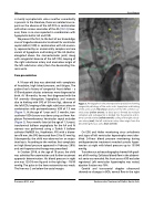
Figure 1. Aortography in the anteroposterior projection showing congenital pathology of the aortic arch: hypoplasia and kinking of the aortic arch (red arrow); absence of the left common ca- rotid artery (yellow arrows); its distal part is formed by multiple collaterals and subsequently is divided into the external and in- ternal carotid arteries (yellow asterisk); coiling of the right subcla- vian artery (oval); the left subclavian artery takes origin from the descending thoracic aorta (blue asterisk).
On ECG and Holter monitoring sinus arrhythmia and signs of left ventricular hypertrophy were iden- tified. 24-hour blood pressure monitoring during antihypertensive treatment revealed systolic hyper- tension at night with blood pressure up to 137/80 mmHg.
Transthoracic echocardiography showed AA gradi- ent of 85 mmHg. Collateral blood flow in the abdomi- nal aorta was revealed. No shunt across VSD occluder registered. Left ventricular hypertrophy was noted, ejection fraction was 78%.
Carotid and transcranial doppler ultrasound showed no changes in BCA, normal flow in the right
Pursanov M. G. et al.
Endovascular Treatment of Gothic Aortic Arch
