
Page 46 - Journal of Structural Heart Disease Volume 5, Issue 6
P. 46
Case Report 270
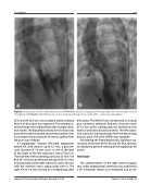
Figure 2. Endovascular closure of perimembranous VSD: Panel a. Left ventriculography showing a large shunt into the right ventricle through the VSD; Panel b. After VSD closure no shunt registered though the occluder VSD – ventricular septal defect.
CCA, the left CCA was not visualized, collateral blood flow in its distal part was registered. The vertebral ar- teries showed normal blood flow with multiple collat- eral vessels. The blood flow velocity in the intracranial parts of the internal carotid and vertebral arteries was not changed. No disturbance of venous outflow from the brain was revealed.
CT-angiography showed left-sided hypoplastic kinked AA, with stenosis up to 6.1 mm, a post-ste- notic diameter of 7.9 mm, and 11.2 mm at the level of the origin of the left subclavian artery (Figure 3). The diameter of the ascending aorta was 25 mm. The BCA (8.7 mm) was divided into the right CCA (7.5 mm) and proximally coiled right subclavian artery (9 mm), with the vertebral artery taking origin from it. The right ICA of 3.4 mm, formed an S-shaped loop after
bifurcation. The left CCA was not detected, in its distal part, numerous collaterals formed a short thin trunk of 3.2 mm, which subsequently was divided into the external and internal carotid arteries. The left subcla- vian artery (8.5 mm) took origin from the descending thoracic aorta. The circle of Willis was complete.
Considering the hemodynamically significant ob- struction at the level of the AA and left CCA absence, we decided to perform stenting of the hypoplastic AA and AI.
Technique
The catheterization of the right internal jugular vein under endotracheal anesthesia was performed, a 5Fr introducer sheath was introduced and an int-
Journal of Structural Heart Disease, December 2019
Volume 5, Issue 6:268-273
