
Page 48 - Journal of Structural Heart Disease Volume 5, Issue 6
P. 48
Case Report 272
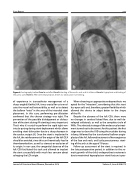
Figure 4. Aortography before Panel a and after Panel b stenting of the aortic arch and its isthmus: Panel a. Hypoplasia and kinking of the aortic arch; Panel b. After stent implantation, there’s no obstruction nor kinking.
of experience in transcatheter management of a sharp-angled (Gothic) AA, it was crucial for us to eval- uate the vessel wall extensibility, as well as to detect the balloon “waist” in the area of the intended stent placement. In this case, performing pre-dilatation confirmed that the chosen strategy was right. The prevention of the possible dislodgement or disloca- tion of the stent during AA stenting is very important. That’s why it’s crucial to perform the rapid right ven- tricular pacing during stent deployment which allows avoiding stent dislocation due to a sharp decrease in the cardiac output [8]. Once the stent is implanted in the AA, the reinforcement of the origin of the left CCA should be avoided, since this can theoretically lead to thromboembolism, as well as stenosis or occlusion of its origin. In our case, the congenital absence of the left CCA facilitated this task and allowed to implant the stent successfully with much less concern about salvaging the LCA origin.
When choosing an appropriate endoprosthesis we opted for the "Intrastent", considering that this stent has open cells and, therefore, greater flexibility which allowed the device to adapt better to the shape of the AA.
Despite the absence of the left CCA, there were no changes in cerebral blood flow, due to well-de- veloped collaterals, as well as the complete circle of Willis. The selected strategy of the endovascular treat- ment turned out to be correct for this patient: the first stage was to close the VSD using the occluder during infancy, followed by the transluminal balloon angio- plasty of the AA, followed treatment of hemangiomas of the face and neck, and lastly percutaneous stent- ing of the arch at the age of 10 years.
Follow up assessment of the stent is required. In the late postoperative period, in addition to the so- matic growth of the child, endoprosthesis restenosis due to neointimal hyperplasia or stent fracture is pos-
Journal of Structural Heart Disease, December 2019
Volume 5, Issue 6:268-273
