
Page 47 - Journal of Structural Heart Disease Volume 5, Issue 6
P. 47
271
Case Report
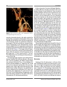
Figure 3. 3D reconstruction (VRT) of CT angiography, demon- strating pathology of the aortic arch.
racardiac electrode placed in the right ventricle for pacing. Next, retrograde catheterization of the aorta through the left femoral artery with the installation of a 9Fr introducer sheath was performed. Unfraction- ated heparin (100 units per kg) was injected intrave- nously. Aortography in the anteroposterior and left anterior oblique (LAO 45o) projections confirmed the congenital anomaly of the Gothic AA with an absence of the left CCA (Figure 1). The length of the AA hypo- plasia was 34 mm with a minimal luminal diameter of 5 mm, while the diameter of the AA proximal to the BCA origin was 11.5 mm, distal to the AI – 9.6 mm. The SPG between the ascending and descending aor- ta was 59 mm Hg.
An exchange length, Amplatz super-stiff guidewire (Boston Scientific-City, MA) was placed in the aortic root. Over this guidewire, 9Fr Mullins sheath (Cook) was advanced to the AA. Then, a Z-med balloon dil- atation catheter (NuMED, Inc., Hopkinton, NY) 12x40 mm and was advanced and placed at the aortic hy- poplasia zone. Under right ventricular pacing at 200 beats/minute, balloon dilatation was performed in order to determine the localization of its “waist” and to predict stent deployment. At a pressure of 7 atm. the “waist” in the inflated balloon disappeared, and its walls were firmly attached to the walls of the aorta. Subsequently, AA stenting was performed.
A 36 mm long stent “Intrastent LD Mega” (Medtron- ic, Fridley, MN) was manually mounted on the same balloon catheter Z-med (12x40 mm). Then, the entire assembly (balloon with the stent mounted on it) was advanced to the area of stenosis. The correct stent position in the AA obstruction area was monitored by contrast injection via the side port of the sheath. When the proper stent position was confirmed, the balloon was manually inflated during rapid right ven- tricular pacing at 200 beats/minute. At a pressure of 10 atm. complete deployment of the stent was achieved with normalization of the AA geometry. Re- peat aortogram showed good result, the stent was fully opened in the AA, with no signs of complications (vessel wall injury) (Figure 4). Repeat pressure gradi- ent revealed the presence of only 6 mmHg across the area. The sheath was removed and hemostasis was achieved by direct pressure. After the operation, the anticoagulant therapy was continued, including intravenous administration of heparin (100 units per kg) twice every 6 hours, followed by oral aspirin at 4 mg/kg per day for 6 months. For the prevention of in- fectious complications, a broad-spectrum antibiotic was administered intraoperatively.
The postoperative period was uneventful. A day af- ter the surgery, the arterial pulse in the lower extrem- ities was distinct, the difference in systolic blood pres- sure between the right hand and leg did not exceed 10 mmHg. Doppler ultrasound after stenting showed restoration of normal blood flow in the abdominal aor- ta, the SPG across the stent was of 20 mmHg. The child was discharged on the 5th day after the procedure. A comprehensive examination with a chest computed tomography was planned at 6 months follow up.
Discussion
Stenting of the AA obstruction is still one of the most challenging endovascular interventions in chil- dren. In this type of congenital heart defect, balloon dilatation is often unsuccessful, and stent implanta- tion is required [15]. Percutaneous interventions for AA hypoplasia are somewhat different from those in coarctation of the aorta [4, 5]. Managing the stenting of the AA and coarctation of the aorta, we have given up performing pre-dilatation because of the risk of aortic wall injury. However, considering the absence
Pursanov M. G. et al.
Endovascular Treatment of Gothic Aortic Arch
