
Page 16 - Journal of Structural Heart Disease - Volume 1 Issue 1
P. 16
Original Research Article 10
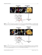
Figure 1a. Paravalvular leak adjacent to an annuloplasty ring. 2D TEE 4-chamber (top row, far right), long axis (top row, center right), and 90-degree views (top row, center left) depict the case of a patient with extensive paravalvular leak spanning across three viewing planes along the posterior portion of the mitral annulus (bottom figure). This is well seen on 3D image (top row far right).
Figure 1b. Paravalvular leak adjacent to a prosthetic valve. 2D TEE long axis (top row, far right), and 90 degree views (top row, center) depict the case of a patient with paravalvular leak spanning across two viewing planes along the anterior wall of the mitral annulus on the side of the aorta and the left atrial appendage (bottom figure showing approximate location of the leak). This is well seen on 3D image (top row far right). Asterisk shows the location of the left atrial appendage.
Structural Heart Disease, May 2015 Volume 1, Issue 1: 9-19
