
Page 18 - Journal of Structural Heart Disease - Volume 1 Issue 1
P. 18
Original Research Article 12
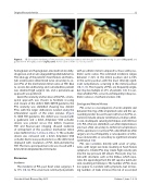
Figure 3. 3D full volume rendering of the mechanical mitral prosthesis with the guide wire in the wrong location (left panel) and guide wire in the right position (right panel). Aortic valve is in the 12 o’clock position.
hemoglobin and haptoglobin, elevated lactate dehy- drogenase, and an unconjugated hyperbilirubinemia. The etiology of the patient’s heart failure and hemo- lytic anemia were determined to be secondary to se- vere PVL of the mechanical mitral valve on TEE. Due to severe deconditioning and comorbidities patient was deemed high surgical risk, and a percutaneous approach was preferred.
Given the anatomy and location of the PVL, a trans- apical approach was chosen to facilitate crossing and closure of the defect. With 3DTEE guidance, the PVL anatomy was identified showing two distinct PVLs with the larger dehiscence located along the inferolateral aspect of the valve annulus (Figure 5). With TEE guidance, the defect was crossed with a guidewire and a 6mm Amplatzer VSD occluder device was placed across the defect, however, TEE and fluoroscopic imaging showed evidence of entrapment of the posterior mechanical mitral valve leaflet (Video 5, Video 6, Video 7). The occluder device was removed and a 4mm Amplatzer VSD occluder device was positioned across the defect and deployed with resolution of PVL demonstrated by TEE. The trans-apical puncture site was closed with a 6mm/4mm Amplatzer Duct Occluder.
Discussion
Incidence
The incidence of PVL post heart valve surgery is 5 to 17% [10-12]. PVL occur more commonly in patients
with prosthetic mitral compared to those with pros- thetic aortic valves. The estimated incidence ranges between 7–32% in the mitral position and 2–10% in the aortic position, with the most clinically signif- icant complications occurring in the mitral position [10,11,13]. The majority of PVLs are frequently single, but may be multiple in 27% of patients [14]. It is un- clear whether PVLs occur more frequently in biopros- thetic valves vs. mechanical valves [10].
Etiology and Natural History
PVL occur as a consequence of an incomplete seal between the ring of the implanted valve and the sur- rounding cardiac tissue. Known risk factors for PVL oc- currence include annular calcification, small prosthet- ic size, inadequate suturing technique, and infection [15]. PVLs that are identified soon after implantations are most often secondary to technical complications of the operation; in contrast, PVLs identified late after surgery are most frequently a consequence of infec- tious endocarditis or secondary to significant annular calcification [16].
PVL size correlates directly with onset of symp- toms; with larger size leaks resulting in heart failure symptoms. Smaller PVL may create high velocity jets into the low pressure left atrium. These jets may col- lide with structures such as the limbus, which sepa- rates, the appendage from the left superior pulmonic vein, resulting in hemolysis. The number of PVLs does not appear to correlate with symptoms, but increas- ing numbers of leaks increase the risk of associated
Structural Heart Disease, May 2015
Volume 1, Issue 1: 9-19
