
Page 19 - Journal of Structural Heart Disease - Volume 1 Issue 1
P. 19
13
Original Research Article
hemolysis [14]. With follow-up, leaks may increase or decrease in size, or, less commonly, may sponta- neously close [13,17,18]. Importantly, the presence of PVL results in turbulent blood flow thereby aug- menting the risk for the development of infective endocarditis in the presence of bacteremia. If regur- gitant flow is significant and not corrected, the natu- ral history of PVLs may mimic that of native valve re- gurgitation. Uncorrected hemolysis eventually results in severe anemia.
Clinical Findings
Patients with symptomatic PVLs present with congestive heart failure in over 90% of cases. Most report NYHA Class III or greater symptoms [19,20]. Clinical presentation may occur immediately after surgery or significantly later [21]. Hemolytic ane- mia is present between 30–75% of cases referred for intervention [19,20].
The regurgitation associated with large PVLs is often associated with a murmur on cardiac ausculta- tion. In para-mitral valve leaks, a blowing, holosystol- ic murmur is typically heard radiating to the axillae. However, paravalvular regurgitatant jets may be ori- ented differently than jets associated with intra-val- vular regurgitation; if the jet is oriented posteriorly, radiation may be noted in the back. If the jet is orient- ed anteriorly, radiation to the base may be heard. The murmur appreciated in para-aortic valve leaks is typi- cally a blowing, decrescendo, diastolic murmur that is heard best at the left sternal border with the patient sitting forward and in end-expiration.
The majority of patients presenting with symptom- atic PVLs have elevations of N-terminal pro-brain na- triuretic peptide. Brain natriuretic peptide is typically elevated in patients with congestive heart failure, but it has also been shown to correlate with the severi- ty and symptoms of aortic and mitral regurgitation [22,23]. When hemolytic anemia is present, laborato- ry studies will show decreased hemoglobin, markedly elevated lactate dehydrogenase, markedly decreased haptoglobin, and increased indirect bilirubin.
Echocardiography
Transesophageal (TEE) and transthoracic (TTE) echocardiography should be used to assess pros-
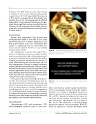
Figure 4.
3D full-volume rendering of the mechanical mitral prosthesiswith6-mmoccluderdeviceinposition.Aorticvalveis in the 12 o’clock position.
Video 4.
expansion.
thetic valve function and the spatial characteristics of PVLs. Color Doppler can help identify the location, direction, and severity of regurgitant blood flow. However, because the spatial resolution of traditional TEE and TTE is limited, the addition of 3D allows for improved spatial resolution and therefore provides more information regarding the size and shape of PVLs [24,25]. This information is especially helpful during percutaneous closure procedures. RT3D TEE allows for operators to visualize the length of the
Case 1: 3D acquisition of 6 mm occluder device
Venturini, J.M. et al.
Real-Time 3D TEE Guidance of Prosthetic Valve PVL
