
Page 20 - Journal of Structural Heart Disease - Volume 1 Issue 1
P. 20
Original Research Article 14
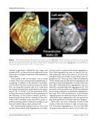
Figure 5. 3D zoomed rendering of the mechanical mitral prosthesis (left panel). With this modality, it is difficult to appreciate the two disks associated with the valve prosthesis in this patient. The paravalvular leaks are not seen. The 3D color zoom acquisition of the mitral prosthesis allows better appreciation of the location and number of paravalvular leaks (right panel). This patient has two paravalvular leaks. Aortic valve is in the 12 o’clock position.
catheter or guidewire, identify the size, shape, and number of PVL, and ensure that any deployed closure device does not impair movement of the mechanical valve leaflets.
The majority of PVL are crescentic, oval, or round in shape. Their track can be parallel, perpendicular, or serpiginous in relation to the direction of prosthet- ic blood flow. The most common location for mitral PVLs are along the posterior wall (5–6 o’clock from the surgeon’s perspective) and along the aortic-mitral curtain (10–11 o’clock) [19,26]. The prevalence of PVLs in the posterior mitral annulus has been attributed to the following: (1) the posterior annulus provides a limited surgical field view for suturing (2) the prox- imity of the circumflex artery may lead to more su- perficial suturing, and (3) calcification and fibrosis are more prevalent in the posterior annulus [24].
The recommended methods used to assess the severity of para-mitral valve regurgitation are similar
to those used to evaluate native mitral regurgitation. Color flow regurgitant jet area, jet density, and sys- tolic pulmonary venous flow reversal are all recom- mended in the assessment of para-mitral valve re- gurgitation [27]. The proportion of the circumference of the sewing ring occupied by the regurgitatant jet provides an approximate guide to severity, with more than 20% indicating severe regurgitation and less than 10% consistent with mild regurgitation [27]. The proximal isovelocity surface area (PISA) measurement has not been validated in paravalvular regurgitation, but large PISA shell measurements of paravalvular regurgitant jets have been reported to be more con- sistent with severe regurgitation [28]. Jet eccentricity may limit traditional assessment with color Doppler.
Aortic PVLs are more commonly located in the vicinity of the non-coronary or right coronary cusps [29]. Para-aortic regurgitation is also assessed with accepted criteria that are used to assess native aor-
Structural Heart Disease, May 2015
Volume 1, Issue 1: 9-19
