
Page 21 - Journal of Structural Heart Disease - Volume 1 Issue 1
P. 21
15
Original Research Article
tic insufficiency. Typical criteria include pressure half- time, jet width, jet density, and diastolic flow reversal in the descending aorta [27].
There are several challenges associated with assess- ment of paravalvular regurgitation with echocardiog- raphy. Mechanical valves create significant image dis- tortion due to acoustic shadowing, and showing may actually hide the presence of regurgitant jets. When multiple PVLs are present, echocardiographic assess- ment is difficult due to eccentric regurgitant jets and absence of validated echocardiographic parameters.
Other Imaging Techniques
PVLs may also be evaluated with EKG-gated com- puted tomographic angiography (CTA). These images can be retrospectively reconstructed to form 4D-re- constructions, that allow for detailed visualization of PVLs. These images have been used to assist plan- ning for percutaneous PVL closure procedures [19]. Like echocardiography, CTA is limited by artifact from high-density structures like the prosthetic valve and extensive calcification. In addition, CTA requires IV contrast and radiation exposure, therefore, the risk of IV contrast and radiation exposure must therefore be weighed against the potential benefit.
Angiography has historically been used to assess the location, size, and hemodynamic severity of PVLs. However, it is difficult to determine the 3D anatomic and spatial characteristics of the defect with angiog- raphy, alone. Invasive assessment of the PVL with test balloons to assess PVL size, distensibility, and hemo- dynamic implications of closure is no longer recom- mended due to the risk of balloon entrapment.
Treatment
Medical therapy in large PVLs is directed at symp- tom reduction by either treating the heart failure or treating the anemia caused by hemolysis. Despite these interventions, the majority of patients with se- vere PVL require definitive, structural correction via either open surgery or transcatheter-based interven- tion.
Until recently, surgical management of PVLs was the only available treatment for severe disease. Sur- gical correction improves overall survival and symp- toms in patients with severe PVL, when compared
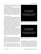
Video 5.
Case 2: Fluoroscopy showing evidence of entrapment of the posterior mechanical mitral valve leaflet.
to medical therapy, alone [12]. Surgery entails either repair of the PVL or re-do replacement of the pros- thetic valve. Many approaches to surgical correction of mitral PVLs have been described, but most involve either direct suturing, patching, or incorporation of autologous tissue from neighboring structures [30- 34]. The choice of repair versus replacement depends
Venturini, J.M. et al.
Real-Time 3D TEE Guidance of Prosthetic Valve PVL
Video 6.
Case 2: 2D TEE showing evidence of entrapment of the posterior mechanical mitral valve leaflet.
