
Page 23 - Journal of Structural Heart Disease - Volume 1 Issue 1
P. 23
17 Original Research Article
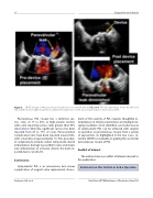
Figure 6. 2D TEE images of the paravalvular leak that was closed with device (left panel). The top right image shows the device in situ and the bottom right image shows diminished significance of this paravalvular leak post device placement.
Percutaneous PVL closure has a technical suc- cess rates of 77 to 88% in high-volume centers, with some reporting success rates greater than 95% [20,25,40,42]. Clinically, significant success has been reported from 67 to 77% of cases. Peri-procedure complication rates have been reported around 10%, with a mortality of approximately 1%. Peri-procedur- al complications include cardiac tamponade, device embolization, damage to prosthetic valve, and stroke. Late embolization of occlusive devices has been re- ported, but is rare [44,45].
Conclusions
Symptomatic PVL is an uncommon, but serious complication of surgical valve replacement. Assess-
ment of the severity of PVL requires thoughtful in- terpretation of clinical presentation and multiple im- aging modalities. Once identified, successful closure of symptomatic PVL can be achieved with surgical re-operation or percutaneous closure from a variety of approaches. As highlighted in the two cases, re- al-time 3D TEE is invaluable in guiding the successful percutaneous closure of PVL.
Conflict of Interest
The authors have no conflict of interest relevant to this publication.
Comment on this Article or Ask a Question
Venturini, J.M. et al.
Real-Time 3D TEE Guidance of Prosthetic Valve PVL
