
Page 22 - Journal of Structural Heart Disease - Volume 1 Issue 1
P. 22
Original Research Article
16
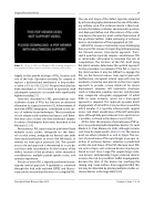
Video 7.
The size and shape of the defect, typically evaluated by echocardiography determines the size of the deliv- ery catheter used. The occlusion device is then load- ed onto the delivery catheter, advanced into position, and deployed. Before and after release of the occlu- sion device, the operator must confirm free motion of the prosthetic leaflets, stable anchoring of occlusion device, and reduction of the regurgitant jet [38].
Mitral PVL closure is technically more challenging than aortic PVL closure. It is typically performed using the femoral venous trans-septal approach. Trans- septal puncture typically requires simultaneous TEE or intracardiac ultrasound to minimize the risk of complication. The location of the PVL itself along the mitral annulus determines the optimal approach for the procedure. For example, if the PVL is close to the atrial septum, it may be difficult to engage the PVL via the femoral venous trans-septal approach. Furthermore, retrograde arterial approach may be needed to snare the wire placed via the trans-septal approach to provide a more stable rail for device deployment. However, left ventricular structures (such as trabeculae, papillary muscles, and chordae) may complicate retrograde engagement of mitral PVRs. In some instances, access via a trans-apical approach is required. This approach provides direct engagement of mitral PVL at any location around the mitral annulus. It is typically achieved with surgical access and direct visualization of the left ventricular apex, although fully percutaneous trans-apical access is possible, as shown in the above cases [39,40].
At this time, the majority of percutaneous PVR re- pairs are performed with Amplatzer devices (St. Jude Medical, St. Paul, Minnesota, USA), although vascular coils have also been used [1,20,25,41-43]. The devices used are either cylindrical or oval in shape. The suc- cess of percutaneous PVL repair hinges on proper se- lection of occlusion devices. Selection is predicated on the size and shape of the PVL. Because most PVL are oval in shape, oval occlusion devices may be pre- ferred in most cases. Large PVL require large occlu- sion devices. Unfortunately, larger occlusion devices increase the risk for prosthetic leaflet impingement, because the discs of the device can overhang the sewing ring. Some authors have suggested that this risk may be alleviated by placing multiple smaller oc- clusive devices in the large defect [38].
Case 2: 2D color Doppler TEE showing evidence of entrapment of the posterior mechanical mitral valve leaflet.
largely on the specific etiology of PVL, location, and size of the leak. Operative mortality for surgery to replace a dysfunctional mechanical or bioprosthet- ic valve is 5% to 14% [35,36]. Hospital mortality has been described as 13% for initial re-operation, with subsequent operations associated with significantly higher mortality [37].
Since first described in 1992, percutaneous tran- scatheter closure of PVLs has become an attractive alternative to surgical correction [3]. Advancement of real-time 3DTEE imaging has contributed to the suc- cess of catheter-based techniques. These procedures do not require cardio-pulmonary bypass, and there- fore may carry a lower risk than traditional surgery. A variety of techniques have been described in the literature [1,3-9].
Percutaneous PVL repair may be performed from multiple access points; retrograde via the femo- ral or radial artery, antegrade via femoral vein (with trans-septal perforation to access the left heart), or directly via trans-apical puncture [38]. The specific access site and approach is determined in a case-by- case basis with consideration for the location of the defect, location of the prosthesis, other anatomical considerations, multiple patient-specific issues, and operator experience.
Closure of aortic PVL is typically performed via ret- rograde arterial approach. A guidewire is advanced through the leak, with real-time 3DTEE and fluoros- copy used to ensure that the wire is crossing the PVL.
Structural Heart Disease, May 2015
Volume 1, Issue 1: 9-19
