
Page 17 - Journal of Structural Heart Disease - Volume 1 Issue 1
P. 17
11
Original Research Article
Video 2.
6mm muscular VSD occluder device (St. Jude Medi- cal, St. Paul, Minnesota, USA) was placed into the de- fect without compromise of leaflet mobility on TEE (Figure 4, Video 4) and fluoroscopy. TEE confirmed closure of the large PVL. Using transthoracic echo- cardiographic guidance, the trans-apical puncture site was closed with a 6mm/4mm Amplatzer Duct Occluder (St. Jude Medical, St. Paul, Minnesota, USA). The patient tolerated the procedure well and was dis- charged home 3 days later.
Case 2
A 72-year-old male with history of rheumatic heart disease requiring mechanical mitral and aortic valve replacements was transferred for consideration of re- peat mitral valve replacement. He was initially hospi- talized with acute pulmonary edema and severe he- molytic anemia requiring blood transfusions. Upon transfer there was significant volume overload, and severe intravascular hemolysis as evidenced by low
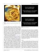
Figure 2. 3D zoom view of the mechanical mitral prosthesis with paravalvular leak as noted. Aortic valve is in the 12 o’clock position.
Association (NYHA) Class III heart failure symptoms and atrial fibrillation. Diagnostic evaluation including right heart catheterization and TEE revealed severe pulmonary hypertension (PA pressure 84/34 mmHg with a mean of 50 mm Hg) and an area of partial dehiscence of the prosthetic mitral valve with a large PVL. Due to her worsening heart failure and history of multiple sternotomies, surgical repair was deemed to be associated with prohibitive risk and a percutaneous approach was planned.
Due to location of the defect (Figure 2, Video 3) and the presence of a mechanical aortic valve, a trans-api- cal approach was chosen for percutaneous closure over a trans-septal or retrograde aortic approach. After apical access was obtained, the paravalvular defect was identified and crossed with a 0.035inch x 150 cm Terumo straight stiff glide wire (Terumo Medical, Somerset, New Jersey, USA) (Figure 3) with 3DTEE guidance. Initially, an 8-mm muscular ventric- ular septal defect occluder (St. Jude Medical, St. Paul, Minnesota, USA) was positioned and deployed into the large defect. On 3DTEE and fluoroscopy, there was evidence of entrapment of a mechanical mitral valve leaflet and the device was retrieved. Next, a
Mechanical mitral prosthetic dehiscence.
Case 1: Mechanical mitral prosthesis with paravalvular leak in the 4 o’clock position.
Video 3.
Venturini, J.M. et al.
Real-Time 3D TEE Guidance of Prosthetic Valve PVL
