
Page 27 - Journal of Structural Heart Disease Volume 2, Issue 1
P. 27
21
Original Research Report
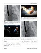
Video 14. The delivery sheath positioned in the left atrium just outside the LSPV. View supplementary video at http://dx.doi. org/10.12945/j.jshd.2016.007.14.vid.14.
Video 15. Corresponding TEE loop showing the sheath in the left atrium near the opening of the LSPV. View supplementary video at http://dx.doi.org/10.12945/j.jshd.2016.007.14.vid.15.
Slide # 35:
Left frame: Crossing the defect with a Judkin’s right coronary artery catheter. The catheter tip is positioned in the left superior pulmonary vein (LSPV) (Video 9).
Video 16. Dilator is removed from the sheath to allow back bleed and prevent air embolism. View supplementary video at http://dx.doi.org/10.12945/j.jshd.2016.007.14.vid.16.
Video 17. Amplatzer septal occluder (ASO) is being passed through the delivery sheath. View supplementary video at http://dx.doi.org/10.12945/j.jshd.2016.007.14.vid.17.
Right frame: A oppy tip Amplatz Supersti TM guide wire (Boston Scienti c, Marlborough, Massa- chusetts, USA) being placed in the LSPV (Video 10).
Jain, S. et al.
Catheter closure of atrial septal defect
