
Page 29 - Journal of Structural Heart Disease Volume 2, Issue 1
P. 29
23
Original Research Report
Video 22.
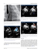
Video 24. TEE loops at 0, con rming adequate capture of all margins before releasing the device from the loading cable. View supplementary video at http://dx.doi.org/10.12945/j. jshd.2016.007.14.vid.24.
Video 25. TEE loops at 45, con rming adequate capture of all margins before releasing the device from the loading cable. View supplementary video at http://dx.doi.org/10.12945/j. jshd.2016.007.14.vid.25.
Slide # 38:
Dilator is removed from the sheath to allow back bleed and prevent air embolism. If the patient is under GA with positive-pressure ventilation, one can back bleed as shown in this movie; but if the patient is breath- ing spontaneously, it is better to back bleed by holding the sheath below the level of the heart in a saline bowl. This helps in preventing inadvertent sucking of air into the sheath resulting in air embolism (Video 16).
Delivery sheath “peeled” back over the loading ca- ble to allow release of the waist and the right atrial disk and de- ployment of device. View supplementary video at http://dx.doi. org/10.12945/j.jshd.2016.007.14.vid.22.
Video 23.
Corresponding TEE loop depicting deployment of the ASO across the defect. View supplementary video at http:// dx.doi.org/10.12945/j.jshd.2016.007.14.vid.23.
Bottom frame: Corresponding TEE loop showing the sheath in the left atrium near the opening of the LSPV (Video 15).
Jain, S. et al.
Catheter closure of atrial septal defect
