
Page 28 - Journal of Structural Heart Disease Volume 2, Issue 1
P. 28
Original Research Report
22
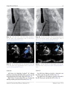
Video 18. Left atrial disk of the ASO being extruded in the left atrium. View supplementary video at http://dx.doi. org/10.12945/j.jshd.2016.007.14.vid.18.
Video 19. Corresponding TEE loop showing left atrial disk in the left atrium. View supplementary video at http://dx.doi. org/10.12945/j.jshd.2016.007.14.vid.19.
Slide # 36:
Left frame: An Amplatzer TorqVueTM 45° delivery sheath (St. Jude, Plymouth, MN, USA) is passed over the Supersti wire into the mouth of the LSPV (Video 11).
Right frame: Corresponding TEE loop showing the delivery sheath positioned in the LSPV (Video 12).
Video 20. The left atrial disk of the ASO being pulled back against the interatrial septum. View supplementary video at http://dx.doi.org/10.12945/j.jshd.2016.007.14.vid.20.
Video 21. Corresponding TEE loop depicting the same. View sup- plementary video at http://dx.doi.org/10.12945/j.jshd.2016.007.14. vid.21.
Slide # 37:
Top left frame: Delivery sheath is advanced over the dilator into the mouth of LSPV (Video 13).
Top right frame: The delivery sheath positioned in the left atrium just outside the LSPV (Video 14).
Journal of Structural Heart Disease, February 2016
Volume 2, Issue 1:15-32
